. 2020 Nov;30(11):6052-6061.
doi: 10.1007/s00330-020-06945-z. Epub 2020 Jun 6.

The Kaiser score reliably excludes malignancy in benign contrast-enhancing lesions classified as BI-RADS 4 on breast MRI high-risk screening exams

Affiliations

The Kaiser score reliably excludes malignancy in benign contrast-enhancing lesions classified as BI-RADS 4 on breast MRI high-risk screening exams

Ruxandra Iulia Milos et al. Eur Radiol. 2020 Nov.

Abstract

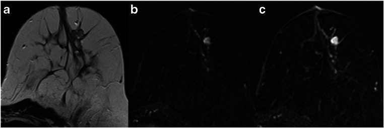
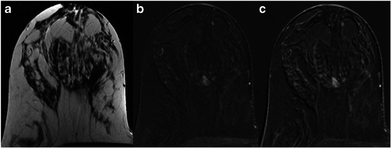
Objectives: MRI is an integral part of breast cancer screening in high-risk patients. We investigated whether the application of the Kaiser score, a clinical decision-support tool, may be used to exclude malignancy in contrast-enhancing lesions classified as BI-RADS 4 on breast MRI screening exams.

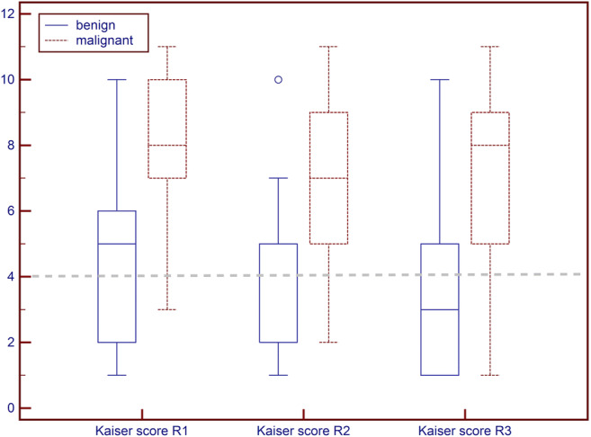
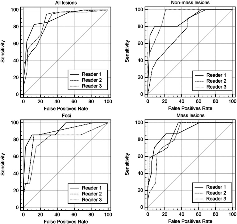
Methods: This retrospective study included 183 consecutive, histologically proven, suspicious (MR BI-RADS 4) lesions detected within our local high-risk screening program. All lesions were evaluated according to the Kaiser score for breast MRI by three readers blinded to the final histopathological diagnosis. The Kaiser score ranges from 1 (lowest, cancer very unlikely) to 11 (highest, cancer very likely) and reflects increasing probabilities of malignancy, with scores greater than 4 requiring biopsy. Receiver operating characteristic (ROC) curve analysis was used to evaluate diagnostic accuracy.

Results: There were 142 benign and 41 malignant lesions, diagnosed in 159 patients (mean age, 43.6 years). Median Kaiser scores ranged between 2 and 5 in benign and 7 and 8 in malignant lesions. For all lesions, the Kaiser score's accuracy, represented by the area under the curve (AUC), ranged between 86.5 and 90.2. The sensitivity of the Kaiser score was high, between 95.1 and 97.6% for all lesions, and was best in mass lesions. Application of the Kaiser score threshold for malignancy (≤ 4) could have potentially avoided 64 (45.1%) to 103 (72.5%) unnecessary biopsies in 142 benign lesions previously classified as BI-RADS 4.

Conclusions: The use of Kaiser score in high-risk MRI screening reliably excludes malignancy in more than 45% of contrast-enhancing lesions classified as BI-RADS 4.

Key points: • The Kaiser score shows high diagnostic accuracy in identifying malignancy in contrast-enhancing lesions in patients undergoing high-risk screening for breast cancer. • The application of the Kaiser score may avoid > 45% of unnecessary breast biopsies in high-risk patients. • The Kaiser score aids decision-making in high-risk breast cancer MRI screening programs.

Keywords: Breast cancer; Clinical; Decision support systems; Magnetic resonance imaging; Screening; Sensitivity and specificity.


Conflict of interest statement

The authors of this manuscript declare no relationships with any companies, whose products or services may be related to the subject matter of the article.

Figures

Fig. 1
Fig. 1
Boxplot of Kaiser score distribution for all three readers (R1, R2, R3) stratified by final diagnosis as benign or malignant. The grey dashed line indicates the biopsy recommendation threshold. It is evident that a majority of benign lesions presents with Kaiser scores below this threshold while most malignant lesions present with Kaiser scores above 4
Fig. 2
Fig. 2
Receiver operating characteristic (ROC) curves for all three readers. All lesions are given in the upper left, non-mass lesions in the upper right, foci in the lower left, and mass lesions in the lower right. Diagnostic performance estimates are summarized in Table 2
Fig. 3
Fig. 3
A 47-year-old high-risk patient: MRI (a T2w; b, c subtracted early and late contrast-enhanced, T1-weighted images) shows a rather circumscribed mass lesion with heterogeneous internal enhancement and wash-out, corresponding to a Kaiser score of 8. Histology revealed a luminal-type invasive lobular cancer, B5b
Fig. 4
Fig. 4
A 35-year-old high-risk patient: MRI (a T2w; b, c subtracted early and late contrast-enhanced, T1-weighted images) shows a circumscribed mass lesion with heterogeneous internal enhancement and wash-out, corresponding to a Kaiser score of 8. Note the hyperintense, fibroadenoma-like T2w-correlate (a). Histology revealed a triple-negative invasive ductal cancer, B5b
Fig. 5
Fig. 5
A 39-year-old high-risk patient: MRI (a T2w; b, c subtracted early and late contrast-enhanced, T1-weighted images) shows a circumscribed mass lesion with heterogeneous internal enhancement and persistent signal increase, corresponding to a Kaiser score of 1. Note the fibroadenoma-like T2w-correlate (a). Histopathology revealed a fibroadenoma, B2
Fig. 6
Fig. 6
A 44-year-old high-risk patient: MRI (a T2w; b, c subtracted early and late contrast-enhanced, T1-weighted images) shows a non-circumscribed, non-mass lesion with heterogeneous internal enhancement and persistent signal increase, corresponding to a Kaiser score of 3. Histopathology revealed benign epithelial proliferations, B2

Similar articles

Cited by

References

    1. Houssami N, Ciatto S, Macaskill P, et al. Accuracy and surgical impact of magnetic resonance imaging in breast cancer staging: systematic review and meta-analysis in detection of multifocal and multicentric cancer. J Clin Oncol. 2008;26:3248–3258. doi: 10.1200/JCO.2007.15.2108. - DOI - PubMed
    1. Pinker K, Helbich TH, Morris EA. The potential of multiparametric MRI of the breast. Br J Radiol. 2017;90:20160715. doi: 10.1259/bjr.20160715. - DOI - PMC - PubMed
    1. Warner E, Messersmith H, Causer P, Eisen A, Shumak R, Plewes D. Systematic review: using magnetic resonance imaging to screen women at high risk for breast cancer. Ann Intern Med. 2008;148:671–679. doi: 10.7326/0003-4819-148-9-200805060-00007. - DOI - PubMed
    1. Bennani-Baiti B, Baltzer PA (2017) MR imaging for diagnosis of malignancy in mammographic microcalcifications: a systematic review and meta-analysis. Radiology 283:692–701 - PubMed
    1. Bennani-Baiti B, Bennani-Baiti N, Baltzer PA. Diagnostic performance of breast magnetic resonance imaging in non-calcified equivocal breast findings: results from a systematic review and meta-analysis. PLoS One. 2016;11:e0160346. doi: 10.1371/journal.pone.0160346. - DOI - PMC - PubMed

MeSH terms