. 2020 Oct:40:101028.
doi: 10.1016/j.molmet.2020.101028. Epub 2020 Jun 3.

Inducible deletion of skeletal muscle AMPKα reveals that AMPK is required for nucleotide balance but dispensable for muscle glucose uptake and fat oxidation during exercise

Affiliations

Inducible deletion of skeletal muscle AMPKα reveals that AMPK is required for nucleotide balance but dispensable for muscle glucose uptake and fat oxidation during exercise

Janne R Hingst et al. Mol Metab. 2020 Oct.

Abstract

Objective: Evidence for AMP-activated protein kinase (AMPK)-mediated regulation of skeletal muscle metabolism during exercise is mainly based on transgenic mouse models with chronic (lifelong) disruption of AMPK function. Findings based on such models are potentially biased by secondary effects related to a chronic lack of AMPK function. To study the direct effect(s) of AMPK on muscle metabolism during exercise, we generated a new mouse model with inducible muscle-specific deletion of AMPKα catalytic subunits in adult mice.

Methods: Tamoxifen-inducible and muscle-specific AMPKα1/α2 double KO mice (AMPKα imdKO) were generated by using the Cre/loxP system, with the Cre under the control of the human skeletal muscle actin (HSA) promoter.

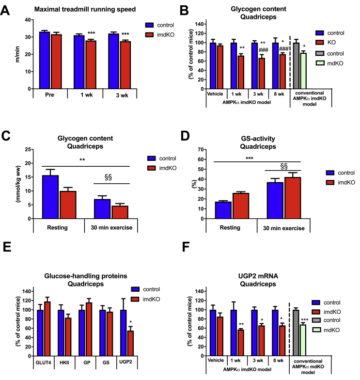
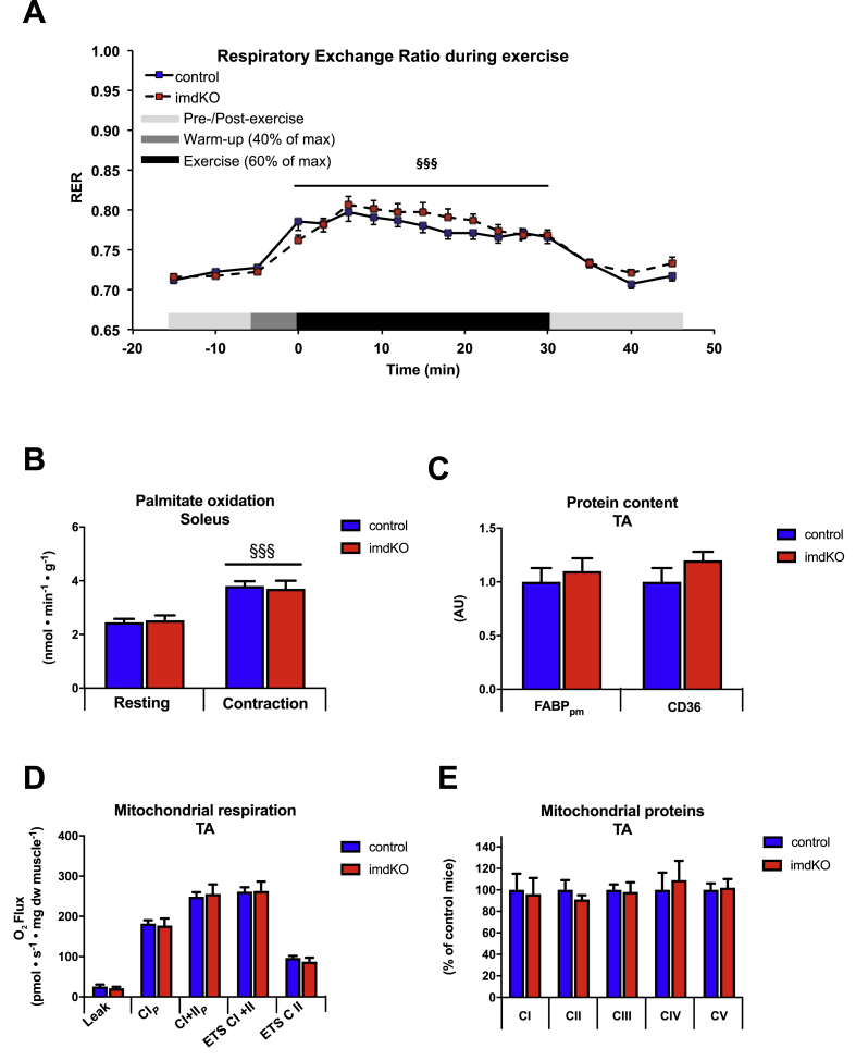
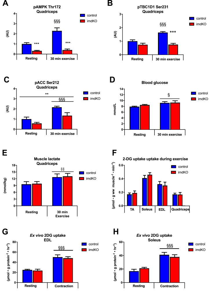
Results: During treadmill running at the same relative exercise intensity, AMPKα imdKO mice showed greater depletion of muscle ATP, which was associated with accumulation of the deamination product IMP. Muscle-specific deletion of AMPKα in adult mice promptly reduced maximal running speed and muscle glycogen content and was associated with reduced expression of UGP2, a key component of the glycogen synthesis pathway. Muscle mitochondrial respiration, whole-body substrate utilization, and muscle glucose uptake and fatty acid (FA) oxidation during muscle contractile activity remained unaffected by muscle-specific deletion of AMPKα subunits in adult mice.

Conclusions: Inducible deletion of AMPKα subunits in adult mice reveals that AMPK is required for maintaining muscle ATP levels and nucleotide balance during exercise but is dispensable for regulating muscle glucose uptake, FA oxidation, and substrate utilization during exercise.

Keywords: AMPK; Exercise; Fat oxidation; Glucose uptake; Glycogen; Muscle metabolism.


Figures

Figure 1
Figure 1
Tamoxifen-induced deletion of AMPKα subunits in adult mice. A–F: Muscle-specific deletion of AMPKα1 and α2 was obtained by expressing a tamoxifen-inducible Cre-recombinase construct driven by the human skeletal muscle actin promotor. The tamoxifen treatment protocol comprised 3 single injections (40 mg/kg bw) separated by 48 h, and mice were investigated 1, 3, and 8 weeks after the last tamoxifen injection. For the vehicle experiment, all mice received injections of sunflower oil. Gene expression of AMPKα1 and α2 subunits was measured in EDL muscle. Protein levels of AMPKα1 and α2 were measured in EDL and heart from control and AMPKα imdKO mice. These data from the AMPKα imdKO model were compared to the conventional AMPKα double KO model (AMPKα mdKO) with chronic lack of AMPK function. Protein levels were measured by immunoblotting, and gene expression was measured by real time PCR and presented relative to TATA-Box Binding Protein (TBP). Data have are normalized to control mice (=100%). Data are given as means ± SEM (n = 5–6 within each group). One-way ANOVA was used for comparing 1, 3, and 8 weeks to vehicle control within AMPKα imdKO mice. An additional t-test was applied to compare AMPKα imdKO with control mice within each time point. ∗p ≤ 0.05, ∗∗p ≤ 0.01, and, ∗∗∗p ≤ 0.001 for difference from corresponding control mice. ##p ≤ 0.01 and ###p ≤ 0.001 for difference from corresponding AMPKα imdKO vehicle. †††p ≤ 0.001 for difference from 1 week AMPKα imdKO.
Figure 2
Figure 2
Normal insulin action despite acute deletion of catalytic AMPK function in skeletal muscle. A–B: 3 weeks after the last tamoxifen injection, control and AMPKα imdKO mice were fasted for 5 h before they were given an intraperitoneal injection of glucose (2 g/kg body weight) dissolved in a 0.9% saline solution. Blood was sampled from the tail vein and analyzed for glucose concentration by a glucometer before (0 min) and 20, 40, 60, 90, and 120 min after injection. Plasma insulin levels were determined at 0, 20, and 40 min by using an insulin ELISA assay (n = 10–12). C: For the insulin tolerance test (ITT), mice were fasted for 2 h, and insulin was injected intraperitoneally (1 U/kg body weight, Actrapid, Novo Nordisk, Bagsværd, Denmark). Tail vein blood glucose concentration was measured before (0 min), 20, 40, and 60 min after injection (n = 10–12). D–E: Isolated EDL and soleus muscles from control and AMPKα imdKO mice were incubated for 30 min in the absence (basal) or presence of 100 μU/ml and 10,000 μU/ml insulin, and muscle glucose uptake was determined by measuring the accumulation of intracellular [3H]-2-deoxyglucose (2DG) (n = 6–8). F–H: Key insulin signaling intermediates in EDL muscle from control and AMPKα imdKO mice were investigated by immunoblotting and are given as representative immunoblots. Data are given as means ± SEM. Two-way RM ANOVA was used to investigate the effect of genotype and time (GTT and ITT) or genotype and insulin concentrations (2DG uptake). ###p ≤ 0.001 for significantly different from basal (0 min). ∗∗p ≤ 0.01 and ∗∗∗p ≤ 0.001 for significantly different compared to basal. §§p ≤ 0.01 and §§§p ≤ 0.001 for significantly different from 100 μU/ml. Line indicates main effect.
Figure 3
Figure 3
Acute deletion of muscle AMPK impairs maximal running speed and reduces muscle glycogen content and UGP2 mRNA. A: Maximal running speed during an incremental running test on a treadmill was assessed in control and AMPKα imdKO mice 1 and 3 weeks after last tamoxifen injection and compared to before tamoxifen treatment (pre; n = 10–20 within each group). B: Muscle glycogen content in quadriceps muscle (normalized to control mice) was measured 1, 3, and 8 weeks after tamoxifen-induced deletion of AMPKα and compared to vehicle control groups (n = 5–6 within each group). C: 3 weeks after last tamoxifen injection, muscle glycogen in quadriceps muscle from control and AMPKα imdKO mice was measured in the rested state and after 30 min of treadmill exercise at the same relative intensity (n = 8–13). D: Glycogen synthase activity was measured as fractional activity in the presence of 0.2 mM G6P and given relative to saturated conditions (8 mM G6P) (n = 8–13). E: Protein levels of GLUT4, HKII, GP, GS, and UGP2 in quadriceps muscle were measured by immunoblotting in control and AMPKα imdKO mice 3 weeks after last tamoxifen injection (n = 5–6). UGP2 mRNA content in quadriceps muscle was determined 1, 3, and 8 weeks after the last tamoxifen injection and compared to the vehicle group (sunflower oil; n = 5–6). One-way ANOVA was used for comparing 1, 3, and 8 weeks to vehicle control within AMPKα imdKO mice. An additional t-test was applied to compare AMPKα imdKO with control mice within each time point. The effect of exercise was investigated by a two-way ANOVA (C and D). Data are given as means ± SEM. ∗p ≤ 0.05, ∗∗p ≤ 0.01, and, ∗∗∗p ≤ 0.001 for effect of genotype within a time point. ###p ≤ 0.001 for difference from vehicle in AMPKα imdKO mice. §§p ≤ 0.01 for main effect of exercise. Line indicates main effect.
Figure 4
Figure 4
Regulation of myocellular nucleotide pool during exercise. Overview of cellular processes regulating cellular nucleotide balance. The increasing ATP utilization during exercise leads to ATP regeneration through the adenylate kinase reaction (2 ADP→ AMP + ATP), which increases accumulation of AMP. To avoid a large accumulation of AMP in the cell, AMP is deaminated to IMP through the enzyme AMP deaminase (AMPD). Intracellular IMP, formed during exercise, can be degraded to inosine (INO) and hypoxanthine (HX), which can leave the muscle cell, potentially causing a nucleotide loss. cN-II: Cytosolic nucleotidase II, cN-IA: Cytosolic nucleotidase IA, PNP: Purine nucleoside phosphorylase, Pi: Inorganic phosphate, NH3: Ammonia.
Figure 5
Figure 5
AMPK is necessary to maintain the cellular nucleotide pool during exercise. A–G: Control and AMPKα imdKO mice performed 30 min of treadmill exercise at the same relative intensity and were compared to corresponding resting mice. Concentration of adenosine triphosphate (ATP), adenosine diphosphate (ADP), adenosine monophosphate (AMP), inosine monophosphate (IMP), hypoxanthine (HX), adenosine (ADO), and inosine (INO) were measured in quadriceps muscle (n = 6–8). H–I: AMPD activity and AMPD1 protein content were measured in quadriceps muscle (n = 6–8). Data are given as means ± SEM. Two-way ANOVA was used for statistical analyses of genotype and exercise. ∗∗p ≤ 0.01 and ∗∗∗p ≤ 0.001 for significant effect of genotype. §§p ≤ 0.01 and §§§p ≤ 0.001 for difference from resting. Line indicates main effect.
Figure 6
Figure 6
AMPK is dispensable for regulation of muscle substrate utilization and mitochondrial function. A: RER before, during, and after 30 min of a single treadmill exercise at approximately 60% of individual maximal running speed (n = 18–20). B: Palmitate oxidation was measured ex vivo in resting or contracting soleus muscles from control and AMPKα imdKO mice (n = 15–18). C. Protein levels of plasma membrane fatty acid binding protein (FABPpm) and cluster of differentiation (CD) 36 were analyzed in TA muscle by immunoblotting (n = 8–13). D: Mitochondrial respiration rates were measured during cumulative addition of substrates in permeabilized TA fibers (n = 9–12). Abbreviations: CIP: Maximal complex I respiration, CI+IIP: Maximal complex I+II linked respiration (capacity for oxidative phosphorylation), ETS (CI+II): Electron transport system capacity (uncoupled respiration) through complex I and II, ETS (CII): Electron transport system capacity through complex II. E: Protein levels of mitochondrial subunits for complex I, II, III, IV, and V in TA muscle were determined by immunoblotting (n = 17–18). Data are given as means ± SEM. The effect of exercise and genotype was investigated by two-way ANOVA. §§§p ≤ 0.001 for difference from resting. Line indicates main effect.
Figure 7
Figure 7
AMPK is not required for exercise and contraction-stimulated glucose uptake in skeletal muscle. A–C: Control and AMPKα imdKO mice were either rested or performed 30 min treadmill exercise at the same relative intensity. Phosphorylation of AMPKα Thr172, TBC1D1 Ser231, and ACC Ser212 was determined in quadriceps muscle by immunoblotting (n = 8–13). D–E: Blood glucose concentration and muscle lactate concentration in quadriceps muscle measured under resting conditions and after 30 min of treadmill exercise (n = 8–13). F: Muscle glucose uptake during 30 min of treadmill exercise was measured in TA, soleus, EDL, and quadriceps muscle from control and AMPKα imdKO mice (n = 8–13). G–H: Isolated EDL and soleus muscle from control and AMPKα imdKO mice were electrically forced to contract, and glucose uptake was measured in resting and contracting muscles (n = 4–8). Data are given as means ± SEM. The effect of genotype and exercise/muscle contraction was investigated by two-way ANOVA. ∗∗p ≤ 0.01 and ∗∗∗p ≤ 0.001 for significant effect of genotype. §p ≤ 0.05, §§p ≤ 0.01 and §§§p ≤ 0.001 for difference from resting. Line indicates main effects.

References

    1. Holloszy J.O., Kohrt W.M. Regulation of carbohydrate and fat metabolism during and after exercise. Annual Review of Nutrition. 1996;16:121–138. - PubMed
    1. Richter E.A., Ruderman N.B. AMPK and the biochemistry of exercise: implications for human health and disease. Biochemical Journal. 2009;418(2):261–275. doi: 10.1042/BJ20082055. - DOI - PMC - PubMed
    1. Hardie D.G., Sakamoto K. AMPK: a key sensor of fuel and energy status in skeletal muscle. Physiology. 2006;21:48–60. doi: 10.1152/physiol.00044.2005. - DOI - PubMed
    1. Corton J.M., Gillespie J.G., Hardie D.G. Role of the AMP-activated protein kinase in the cellular stress response. Current Biology: CB. 1994;4(4):315–324. doi: 10.1016/S0960-9822(00)00070-1. - DOI - PubMed
    1. Winder W.W., Hardie D.G. AMP-activated protein kinase, a metabolic master switch: possible roles in Type 2 diabetes. American Journal of Physiology - Endocrinology And Metabolism. 1999;277(1):E1–E10. doi: 10.1152/ajpendo.1999.277.1.e1. - DOI - PubMed

Publication types

MeSH terms