. 2020 Jul 24;295(30):10394-10405.
doi: 10.1074/jbc.RA119.010984. Epub 2020 Jun 5.

A substitution in cGMP-dependent protein kinase 1 associated with aortic disease induces an active conformation in the absence of cGMP

Affiliations

A substitution in cGMP-dependent protein kinase 1 associated with aortic disease induces an active conformation in the absence of cGMP

Matthew H Chan et al. J Biol Chem. .

Abstract

Type 1 cGMP-dependent protein kinases (PKGs) play important roles in human cardiovascular physiology, regulating vascular tone and smooth-muscle cell phenotype. A mutation in the human PRKG1 gene encoding cGMP-dependent protein kinase 1 (PKG1) leads to thoracic aortic aneurysms and dissections. The mutation causes an arginine-to-glutamine (RQ) substitution within the first cGMP-binding pocket in PKG1. This substitution disrupts cGMP binding to the pocket, but it also unexpectedly causes PKG1 to have high activity in the absence of cGMP via an unknown mechanism. Here, we identified the molecular mechanism whereby the RQ mutation increases basal kinase activity in the human PKG1α and PKG1β isoforms. Although we found that the RQ substitution (R177Q in PKG1α and R192Q in PKG1β) increases PKG1α and PKG1β autophosphorylation in vitro, we did not detect increased autophosphorylation of the PKG1α or PKG1β RQ variant isolated from transiently transfected 293T cells, indicating that increased basal activity of the RQ variants in cells was not driven by PKG1 autophosphorylation. Replacement of Arg-177 in PKG1α with alanine or methionine also increased basal activity. PKG1 exists as a parallel homodimer linked by an N-terminal leucine zipper, and we show that the WT chain in WT-RQ heterodimers partly reduces basal activity of the RQ chain. Using hydrogen/deuterium-exchange MS, we found that the RQ substitution causes PKG1β to adopt an active conformation in the absence of cGMP, similar to that of cGMP-bound WT enzyme. We conclude that the RQ substitution in PKG1 increases its basal activity by disrupting the formation of an inactive conformation.

Keywords: autophosphorylation; cardiovascular system; cyclic GMP (cGMP); enzyme structure; hydrogen exchange mass spectrometry; hydrogen/deuterium exchange; kinase signaling; mutagenesis; mutant; protein kinase G (PKG); thoracic aortic aneurysm and dissection (TAAD).


Conflict of interest statement

Conflict of interest—The authors declare no conflicts of interest with the content of this article.

Figures
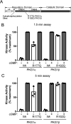
Figure 1.
Figure 1.
In vitro kinase activity of PKG1α R177Q and PKG1β R192Q. A, domain organization of PKG1 highlighting the locations of autophosphorylation sites and the activating RQ mutation. The regulatory domain contains leucine zipper (LZ), autoinhibitory (AI), and cyclic nucleotide binding (CNB-A and CNB-B) subdomains. B and C, kinase assays were performed using purified PKG1α and PKG1β as described in Experimental procedures. Reactions were stopped after 1.5 min (B) or 5 min (C). Data are from three independent protein preparations, with each point representing the average of three kinase reactions for each preparation. Bars show means ± S.D., n = 3.
Figure 2.
Figure 2.
The RQ mutation causes higher autophosphorylation in vitro but not in cells. A, in vitro PKG1α autophosphorylation. Purified PKG1α was incubated for 5 min with 32PO4–γ-ATP under reaction conditions identical to those used for in vitro kinase assays (in the absence of peptide substrate). Phosphate incorporation was determined by SDS-PAGE/autoradiography. Equal loading of the kinase is shown by Western blotting with an anti-Flag antibody. B, performed as in panel A, except using purified PKG1β. C, 293T cells were transfected with expression vectors for Flag-tagged WT and R177Q PKG1α. Six hours posttransfection, cells were incubated with 32PO4 for three hours, and then some cells were treated with 8-pCPT–cGMP for one hour. Upper, PKG was isolated by anti-FLAG immunoprecipitation, and phosphate incorporation was analyzed by SDS-PAGE/autoradiography. Lower, equal PKG amounts were determined by anti-Flag Western blots. D, autophosphorylation of PKG1β in 293T cells performed as in panel C.
Figure 3.
Figure 3.
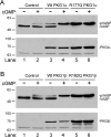
The RQ mutation increases PKG kinase activity in intact cells. A, 293T cells were cotransfected with expression vectors for Myc-tagged VASP and either WT or R177Q PKG1α. At 24 h posttransfection, some cells were treated with 100 μm 8-pCPT–cGMP for one hour. Cell lysates were analyzed by SDS-PAGE/immunoblotting using antibodies recognizing Myc-epitope (upper) or PKG1 (lower). B, experiment was performed as in panel A, but VASP was cotransfected with WT or R192Q PKG1β. The gel shift of VASP indicates S239 phosphorylation.
Figure 4.
Figure 4.
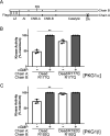
Interchain communication regulates RQ-PKG1 activity. A, PKG1 domain map showing dimerized active and dead PKG chains. RQ indicates the site of the activating mutation in CNB-A, and DA indicates mutation of the catalytic aspartic acid to alanine, which causes a loss of kinase activity. B and C, 293T cells were cotransfected with Flag-tagged dead PKG1, with or without the RQ mutation, and untagged active RQ-mutant PKG1. In vitro kinase reactions were stopped at 1.5 min to measure the activity of untagged PKG1α R177Q (B) or PKG1β R192Q (C). Data are from three independent protein preparations, with each point representing the average from three kinase reactions for each preparation. Bars show means ± S.D., n = 3. **, p > 0.01 by two-tailed Student's t test.
Figure 5.
Figure 5.
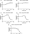
Effects of cyclic nucleotide analog inhibitors on RQ PKG1 activity. In vitro basal kinase activity of purified R177Q PKG1α or R192Q PKG1β measured in the presence of increasing concentrations of Rp-8-pCPT–cGMPS (A and B) or Rp-8-pCPT-PET–cGMPS (C and D). E, In vitro kinase activity of isolated PKG1 catalytic domain (CD) measured in the presence of increasing Rp-8-pCPT-PET-cGMPS concentrations. For all reactions, basal activity in the absence of inhibitor was set at 100%. Data are the average from three experiments using three independent protein preparations. Bars show means ± S.D., n = 3.
Figure 6.
Figure 6.
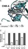
Mutation of R177 to Ala, Met, or Gln leads to increased basal kinase activity. A, molecular model of inactive PKG1α showing putative packing of PKGα R177 in an inactive conformation. B, in silico-predicted destabilizing effects of mutations at R177 using the DUET server (ΔΔG in kcal/mol). C, kinase assays comparing basal to maximum cGMP-stimulated activation of WT and mutant PKG1α. Data are from triplicate reactions from a single protein preparation. The experiment was performed twice with similar results.
Figure 7.
Figure 7.
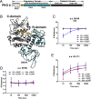
H/D exchange in inhibitory loop residues. A, PKG1β domain organization colored according to the molecular model shown in panel B. LZ, leucine zipper; AI, autoinhibitory loop; CNB-A and CNB-B, cyclic nucleotide binding pockets; and catalytic, the catalytic domain. (B) Molecular model of PKG1β in an inhibited conformation with the regulatory domain CNBs colored teal, catalytic domain in gray, and amino acids 69–86 of the linker/autoinhibitory loop colored yellow. (C) Time-dependent H/D exchange in residues 69–86 (AI), which bind within the catalytic cleft. (D) Time-dependent H/D exchange in residues 59–68, which are N terminal to the autoinhibitory loop. (E) Time-dependent H/D exchange in residues 93–111, which are C terminal to the autoinhibitory loop. Wt, WT PKGIβ; Mut, R192Q PKGIβ. Graphs show the number of deuterons incorporated into the peptides as a function of time. H/D exchange data are averages from two independent H/D exchange reactions performed with two separate protein preparations.
Figure 8.
Figure 8.
H/D exchange in the catalytic cleft of WT and R192Q PKG1β. H/D exchange profiles of peptides from WT (Wt) and R192Q (Mut) PKG1β in the presence or absence of cGMP are shown mapped to a molecular model of inactive PKG1β. The regulatory domain is colored teal and the catalytic domain is mainly colored gray, with the following exceptions: the region containing amino acids 521–533 is colored green; 547–556 is colored red; and 565–583 is colored dark blue. Graphs show the number of deuterons incorporated into the peptides as a function of time. H/D exchange data are the averages from two independent H/D exchange reactions performed with separate protein preparations.

Similar articles

Cited by

References

    1. Hofmann F., Bernhard D., Lukowski R., and Weinmeister P. (2009) cGMP regulated protein kinases (cGK). Handb. Exp. Pharmacol. 191, 137–162 10.1007/978-3-540-68964-5_8 - DOI - PubMed
    1. Francis S. H., Busch J. L., Corbin J. D., and Sibley D. (2010) cGMP-dependent protein kinases and cGMP phosphodiesterases in nitric oxide and cGMP action. Pharmacol. Rev. 62, 525–563 10.1124/pr.110.002907 - DOI - PMC - PubMed
    1. Friebe A., Sandner P., and Schmidtko A. (2017) Meeting report of the 8th International Conference on cGMP “cGMP: generators, effectors, and therapeutic implications” at Bamberg, Germany, from June 23 to 25, 2017. Naunyn. Schmiedebergs Arch. Pharmacol. 390, 1177–1188 10.1007/s00210-017-1429-5 - DOI - PMC - PubMed
    1. Casteel D. E., Zhuang S., Gudi T., Tang J., Vuica M., Desiderio S., and Pilz R. B. (2002) cGMP-dependent protein kinase I beta physically and functionally interacts with the transcriptional regulator TFII-I. J. Biol. Chem. 277, 32003–32014 10.1074/jbc.M112332200 - DOI - PubMed
    1. Kato M., Blanton R., Wang G. R., Judson T. J., Abe Y., Myoishi M., Karas R. H., and Mendelsohn M. E. (2012) Direct binding and regulation of RhoA protein by cyclic GMP-dependent protein kinase Ialpha. J. Biol. Chem. 287, 41342–41351 10.1074/jbc.M112.421040 - DOI - PMC - PubMed

Publication types

MeSH terms

Substances

Associated data