

Interobserver variation in the classification of thymic lesions including biopsies and resection specimens in an international digital microscopy panel
- PMID: 32506527
- PMCID: PMC7702114
- DOI: 10.1111/his.14167
Interobserver variation in the classification of thymic lesions including biopsies and resection specimens in an international digital microscopy panel
Abstract
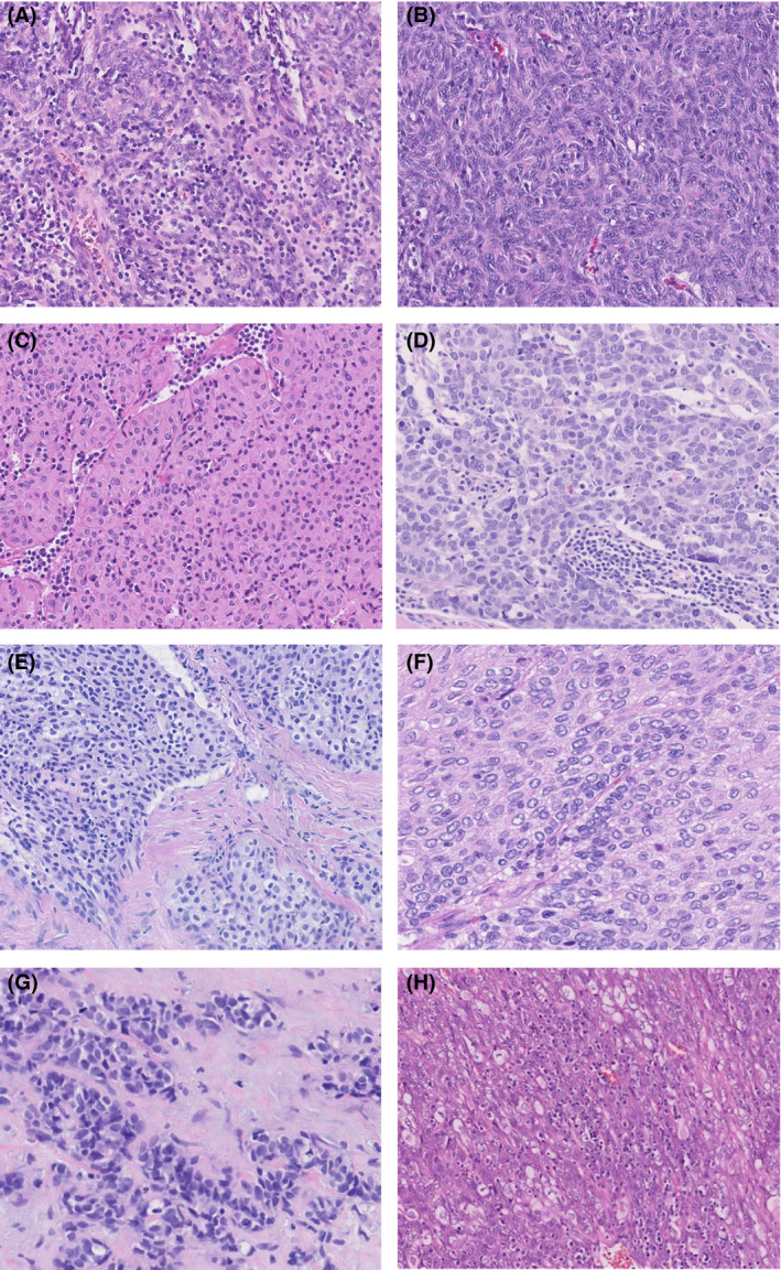
Aims: Thymic tumours are rare in routine pathology practice. Although the World Health Organization (WHO) classification describes a number of well-defined categories, the classification remains challenging. The aim of this study was to investigate the reproducibility of the WHO classification among a large group of international pathologists with expertise in thymic pathology and by using whole slide imaging to facilitate rapid diagnostic turnover.
Methods and results: Three hundred and five tumours, consisting of 90 biopsies and 215 resection specimens, were reviewed with a panel-based virtual microscopy approach by a group of 13 pathologists with expertise in thymic tumours over a period of 6 years. The specimens were classified according to the WHO 2015 classification. The data were subjected to statistical analysis, and interobserver concordance (Fleiss kappa) was calculated. All cases were diagnosed within a time frame of 2 weeks. The overall level of agreement was substantial (κ = 0.6762), and differed slightly between resection specimens (κ = 0.7281) and biopsies (κ = 0.5955). When analysis was limited to thymomas only, and they were grouped according to the European Society for Medical Oncology Clinical Practice Guidelines into B2, B3 versus A, AB, B1 and B3 versus A, AB, B1, B2, the level of agreement decreased slightly (κ = 0.5506 and κ = 0.4929, respectively). Difficulties arose in distinguishing thymoma from thymic carcinoma. Within the thymoma subgroup, difficulties in distinction were seen within the B group.
Conclusions: Agreement in diagnosing thymic lesions is substantial when they are assessed by pathologists with experience of these rare tumours. Digital pathology decreases the turnaround time and facilitates access to what is essentially a multinational resource. This platform provides a template for dealing with rare tumours for which expertise is sparse.
Keywords: interobserver variation; thymoma; tumour classification; whole slide imaging.
© 2020 The Authors. Histopathology published by John Wiley & Sons Ltd.
Conflict of interest statement
The Author(s) declare(s) that there is no conflict of interest.
Figures
References
-
- de Jong WK, Blaauwgeers JL, Schaapveld M, Timens W, Klinkenberg TJ, Groen HJ. Thymic epithelial tumours: a population‐based study of the incidence, diagnostic procedures and therapy. Eur. J. Cancer 2008; 44; 123–130. - PubMed
-
- Marx A, Ströbel P, Badve SS et al ITMIG Consensus Statement on the Use of the WHO Histological Classification of Thymoma and Thymic Carcinoma: refined definitions, histological criteria and reporting. J. Thorac. Oncol. 2014; 9; 596–611. - PubMed
-
- Travis WD, Brambilla E, Burke AP, Marx A, Nicholson AG, eds. World Health Organization classification of tumours of the lung, pleura, thymus and heart. 4th ed. Lyon: IARC Press, 2015. - PubMed
-
- Bernatz PE, Harrison EG, Clagett OT. Thymoma: a clinicopathologic study. J. Thorac. Cardiovasc. Surg. 1961; 42; 424–444. - PubMed
MeSH terms
LinkOut - more resources
Full Text Sources
Medical
