. 2020 Sep;111(9):3338-3349.
doi: 10.1111/cas.14516. Epub 2020 Jul 5.

RNA sequencing of plasma exosomes revealed novel functional long noncoding RNAs in hepatocellular carcinoma

Affiliations

RNA sequencing of plasma exosomes revealed novel functional long noncoding RNAs in hepatocellular carcinoma

Xuejing Huang et al. Cancer Sci. 2020 Sep.

Abstract

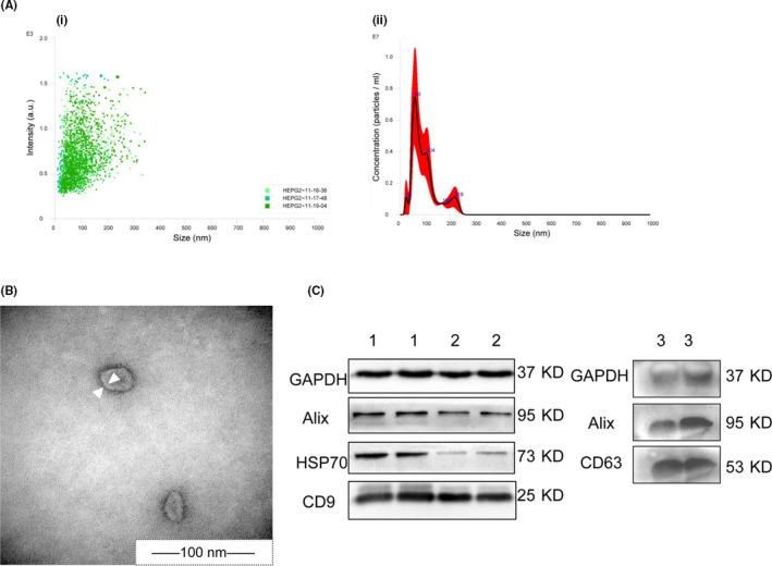
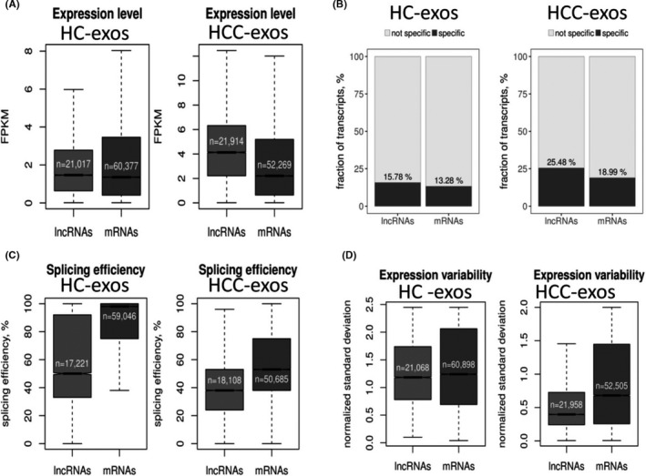
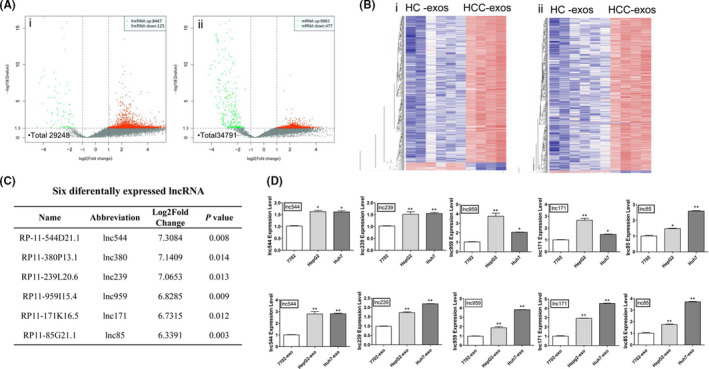
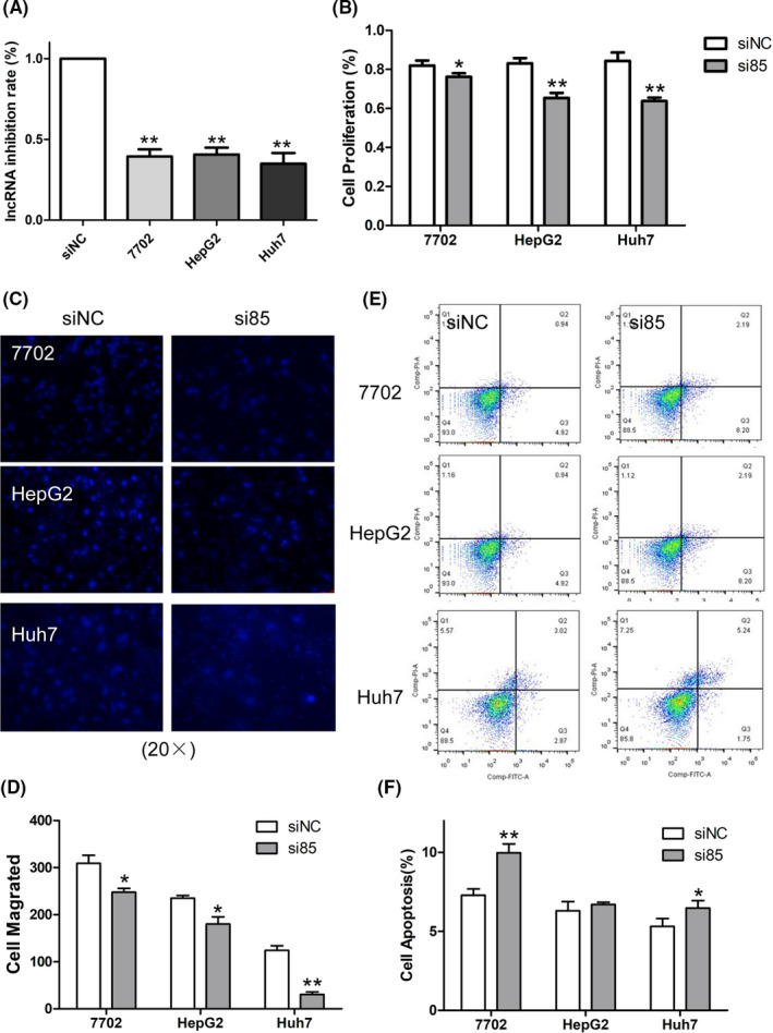
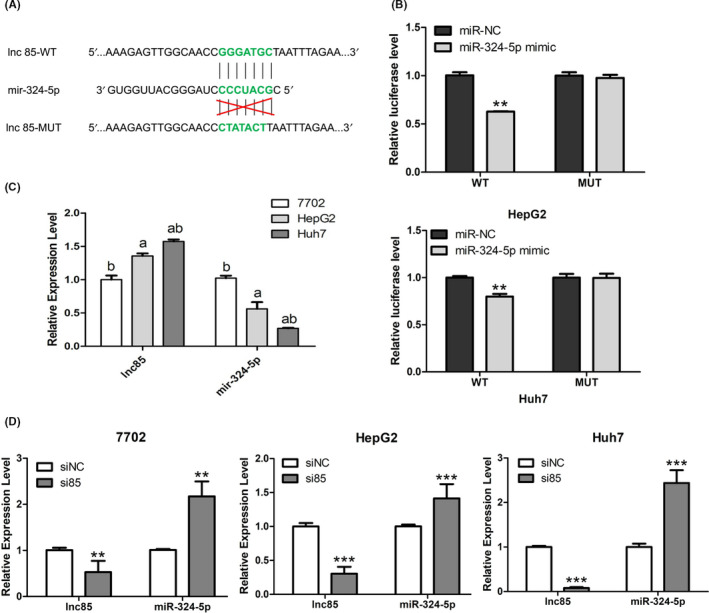
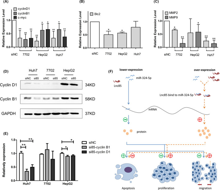
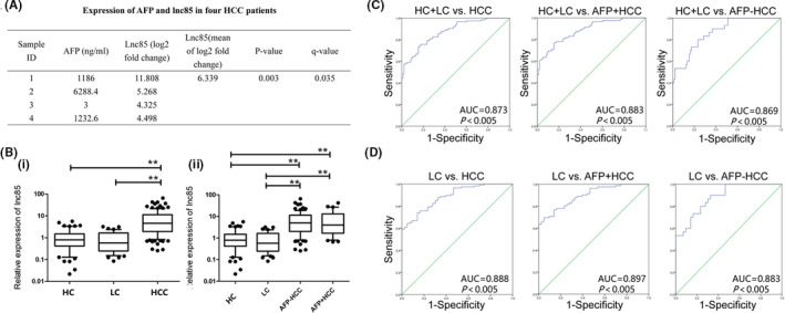
Exosomal long noncoding RNA (lncRNA) has been found to be associated with the development of cancers. However, the expression characteristics and the biological roles of exosomal lncRNAs in hepatocellular carcinoma (HCC) remain unknown. Here, by RNA sequencing, we found 9440 mRNAs and 8572 lncRNAs were differentially expressed (DE-) in plasma exosomes between HCC patients and healthy controls. Exosomal DE-lncRNAs displayed higher expression levels and tissue specificity, lower expression variability and splicing efficiency than DE-mRNAs. Six candidate DE-lncRNAs (fold change 6 or more, P ≤ .01) were high in HCC cells and cell exosomes. The knockdown of these candidate DE-lncRNAs significantly affected the migration, proliferation, and apoptosis in HCC cells. In particular, a novel DE-lncRNA, RP11-85G21.1 (lnc85), promoted HCC cellular proliferation and migration by targeted binding and regulating of miR-324-5p. More importantly, the level of serum lnc85 was highly expressed in both Alpha-fetoprotein (AFP)-positive and AFP-negative HCC patients and allowed distinguishing AFP-negative HCC from healthy control and liver cirrhosis (area under the receiver operating characteristic curve, 0.869; sensitivity, 80.0%; specificity, 76.5%) with high accuracy. Our finding offers a new insight into the association between the dysregulation of exosomal lncRNA and HCC, suggesting that lnc85 could be a potential biomarker of HCC.

Keywords: RP11-85G21.1; hepatocellular carcinoma; long noncoding RNA; miR-324-5p; plasma exosome.


Conflict of interest statement

The authors have no conflict of interest.

Figures

FIGURE 1
FIGURE 1
Identification of exosomes. A, NanoSight of cell exosomes. (i) Three fields of view for analysis. (ii) Intensity curve image of exosome size. B, Transmission electron microscopy images of cell exosomes. Arrowheads indicate the bilayer membrane. C, Western blot images of exosome proteins (exos). (1) HepG2‐exos, (2) HL‐7702‐exos. (3) Plasma‐exos. HSP70, heat shock protein 70
FIGURE 2
FIGURE 2
Expression characteristics of exosomal (exos) long noncoding RNAs (lncRNAs) and mRNAs. A, Median expression levels of lncRNA and mRNA transcripts. B, Specific expression lncRNA and mRNA transcript. C, Median splicing efficiency levels of lncRNA and mRNA transcripts. D, Interindividual variability of differentially expressed (DE)‐lncRNA and DE‐mRNA transcripts
FIGURE 3
FIGURE 3
Differentially expressed long noncoding RNAs (DE‐lncRNAs) and DE‐mRNAs between hepatocellular carcinoma (HCC) and healthy controls (HC). A, Volcano plot showing DE‐lncRNAs (i) and DE‐mRNAs (ii) between HCC exosomes (exos) and HC‐exos. Red and green points represent higher and lower expression of transcription (mRNA or lncRNA) with statistical significance (fold change ≥ or ≤2.0, respectively; P ≤ .05) (X axes, fold change values; Y axes, P values). B, Hierarchical clustering of DE‐lncRNA (i) and DE‐mRNA (ii) between HCC‐exos and HC‐exos (red, high expression; blue, low expression). C, Expression levels of 6 DE‐lncRNAs (fold change ≥ 6.0; P ≤ .05) in HCC‐exos. D, Respective expression level of 5 DE‐lncRNAs in HCC cells and cell exosomes by quantitative real‐time PCR. Data are expressed as mean ± SD of 3 independent experiments. *P ≤ .05, **P ≤ .005 vs. control (7702 and 7702‐exo as control, respectively)
FIGURE 4
FIGURE 4
Knockdown of long noncoding RNA 85 (lnc85) significantly regulated cell proliferation, migration, and apoptosis phenotypes in hepatocellular carcinoma cells. A, Inhibition rate of lnc85 after transfection with siRNA. B, CCK‐8 was used to detect cell proliferation. C, D, Transwell assay was used to detect cell migration (20×) (C) and quantification was carried out (D). E, F, Flow cytometry was used to detect cell apoptosis (E) and quantification was carried out (F). Data are expressed as the mean ± SD of 3 independent experiments. *P ≤ .05, **P ≤ .005 vs. siNC (si‐negative control)
FIGURE 5
FIGURE 5
Targeting relationship between long noncoding RNA 85 (lnc85) and microRNA (miR)‐324‐5p. A, miR‐324‐5p putative binding site on lnc85. B, Relative luciferase activity was detected in HepG2 and Huh7 cells cotransfected with pmirGLO‐lnc85‐WT or pmirGLO‐lnc85‐MUT vectors and mir‐324‐5p mimic or miR‐negative control (miR‐NC). C, D, Expression level of lnc85 and miR‐324‐5p before (C) and after (D) silencing of lnc85 in HL‐7702, HepG2, and Huh7 cells. Data are expressed as the mean ± SD of 3 independent experiments. *P ≤ .05, **P ≤ .005, ***P ≤ .0005 vs. control
FIGURE 6
FIGURE 6
Scarcity of long noncoding RNA 85 (lnc85) enhances the suppression ability of microRNA (miR)‐324‐5p to its downstream gene. A‐C, mRNA expression of proliferation genes (A), antiapoptosis gene (B), and premigration genes (C) is decreased when lnc85 is silenced. D, E, Western blot images (D) and quantification (E) show downregulation of proliferation proteins after silencing of lnc85. F, Working model of lnc85 in hepatocellular carcinoma. Data are expressed as mean ± SD of 3 independent experiments. *P ≤ .05, **P ≤ .005 vs. siNC (si‐negative control)
FIGURE 7
FIGURE 7
Long noncoding RNA 85 (lnc85) as a biomarker of hepatocellular carcinoma (HCC). A, Expression levels of α‐fetoprotein (AFP) and lnc85 in HCC exosomes. B, Lnc85 expression is higher in HCC patients’ serum (i), whether it is AFP‐negative (−) or AFP‐positive (+) (ii), than in healthy controls (HC) and patients with liver cirrhosis (LC). C, Receiver operating characteristic (ROC) curves of lnc85 diagnosis of HC, LC, and HCC patients. D, ROC curves of lnc85 diagnosis of LC and HCC patients. *P ≤ .05, **P ≤ .005 vs. HC

Similar articles

Cited by

References

    1. Venook AP, Papandreou C, Furuse J, de Guevara LL. The incidence and epidemiology of hepatocellular carcinoma: a global and regional perspective. Oncologist. 2010;15(Suppl 4):5‐13. - PubMed
    1. Jemal A, Bray F, Center MM, Ferlay J, Ward E, Forman D. Global cancer statistics. CA Cancer J Clin. 2011;61:69‐90. - PubMed
    1. Chiang JK, Koo M, Kuo TB, Fu CH. Association between cardiovascular autonomic functions and time to death in patients with terminal hepatocellular carcinoma. J Pain Symptom Manage. 2010;39:673‐679. - PubMed
    1. Yang X, Xie X, Xiao YF, et al. The emergence of long non‐coding RNAs in the tumorigenesis of hepatocellular carcinoma. Cancer Lett. 2015;360:119‐124. - PubMed
    1. Chen JG, Zhang SW. Liver cancer epidemic in China: past, present and future. Semin Cancer Biol. 2011;21:59‐69. - PubMed

MeSH terms