. 2020 Dec;84(6):3054-3070.
doi: 10.1002/mrm.28338. Epub 2020 Jun 7.

Advancing machine learning for MR image reconstruction with an open competition: Overview of the 2019 fastMRI challenge

Affiliations

Advancing machine learning for MR image reconstruction with an open competition: Overview of the 2019 fastMRI challenge

Florian Knoll et al. Magn Reson Med. 2020 Dec.

Abstract

Purpose: To advance research in the field of machine learning for MR image reconstruction with an open challenge.

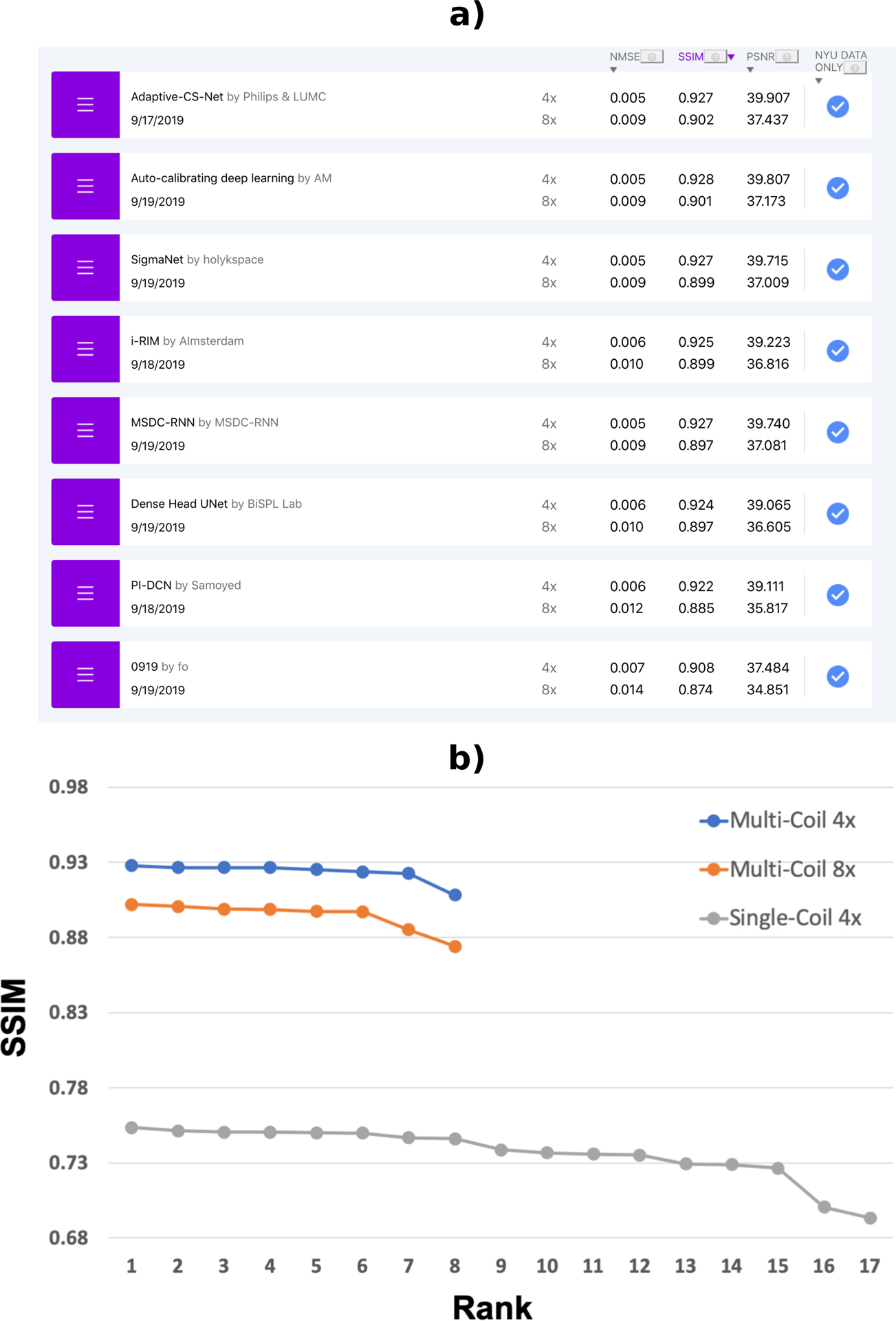
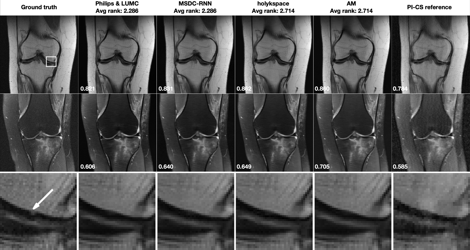
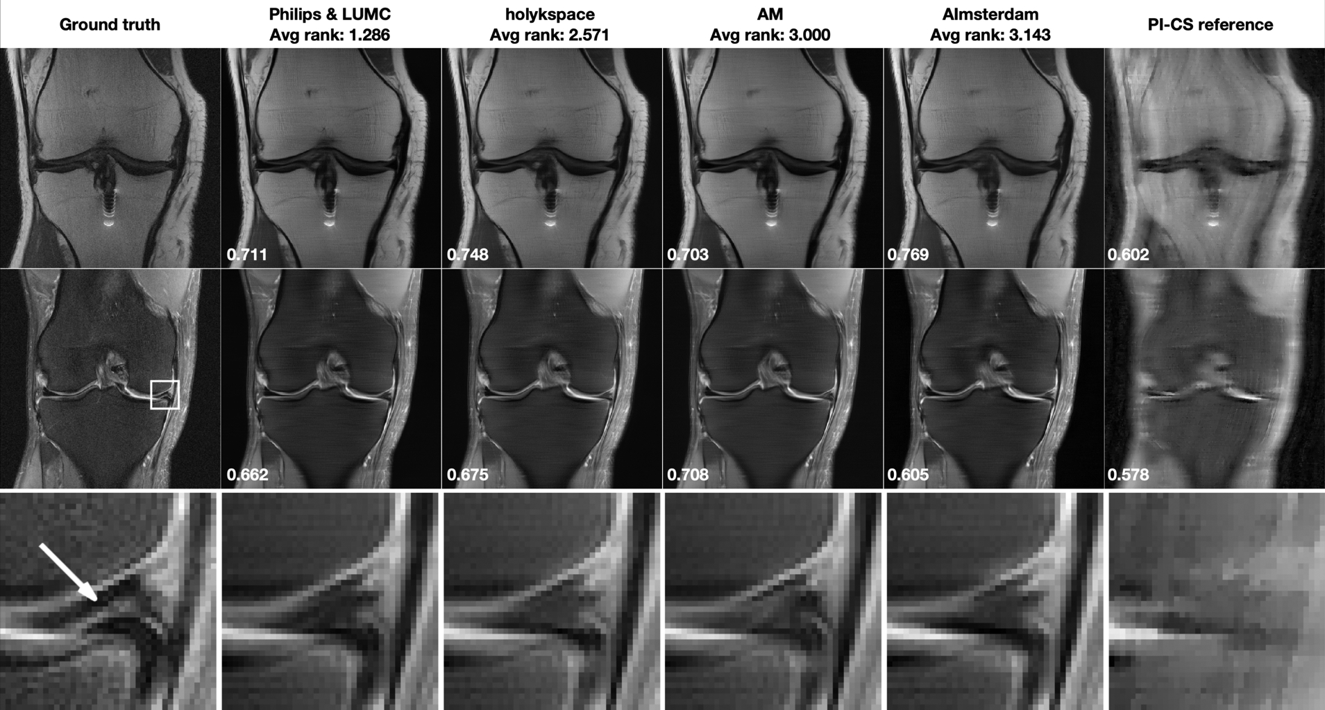
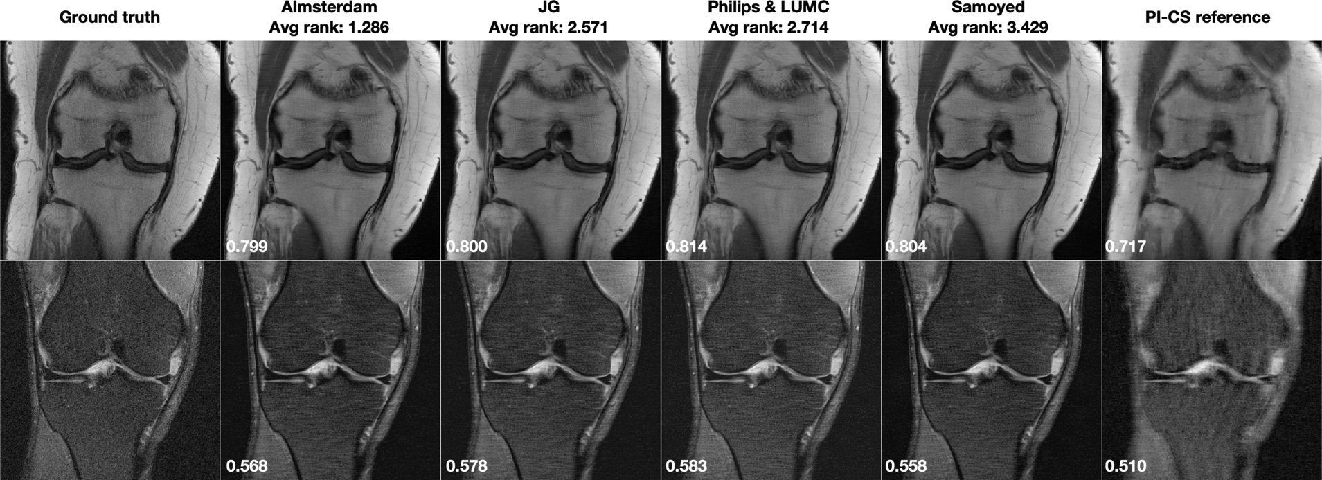
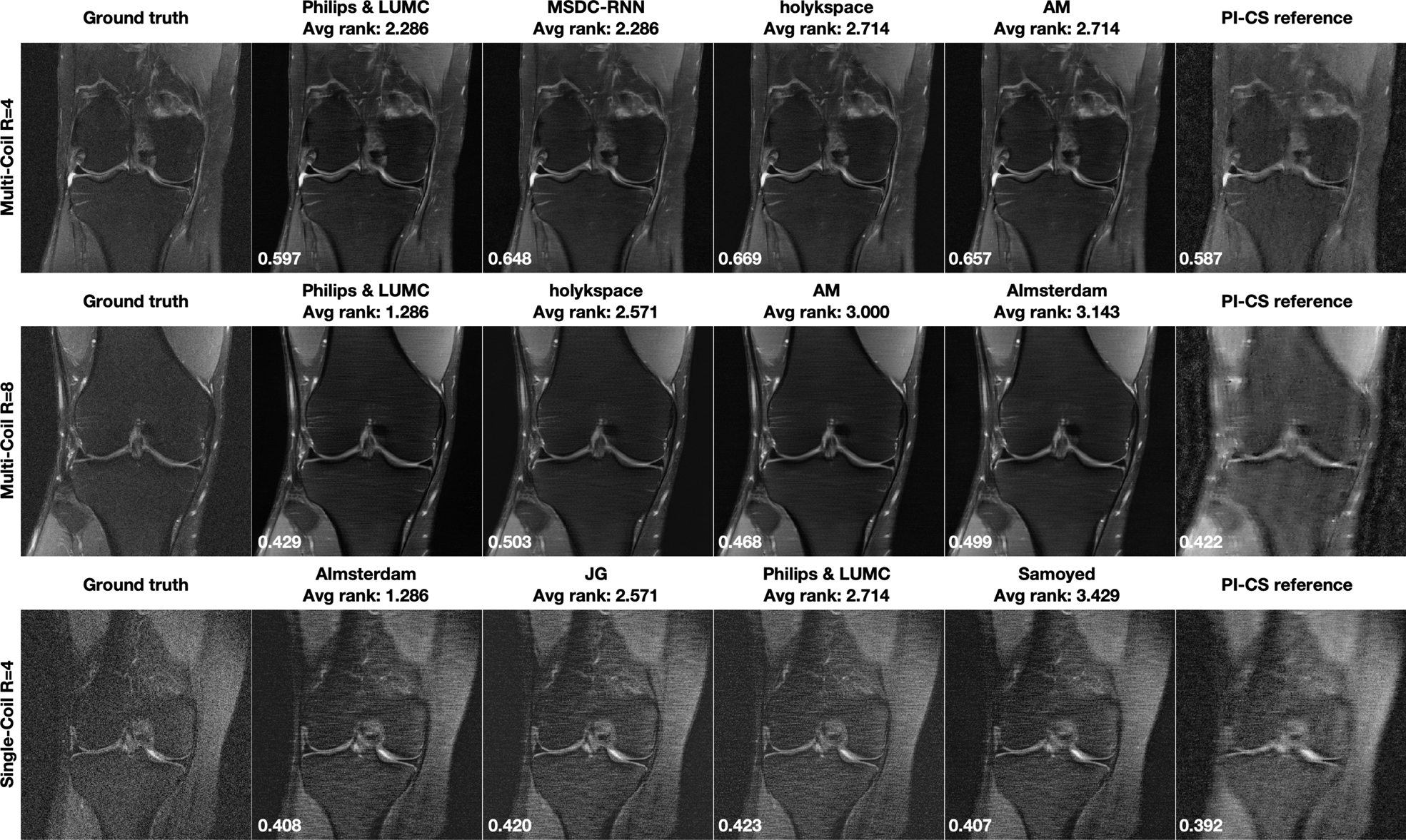
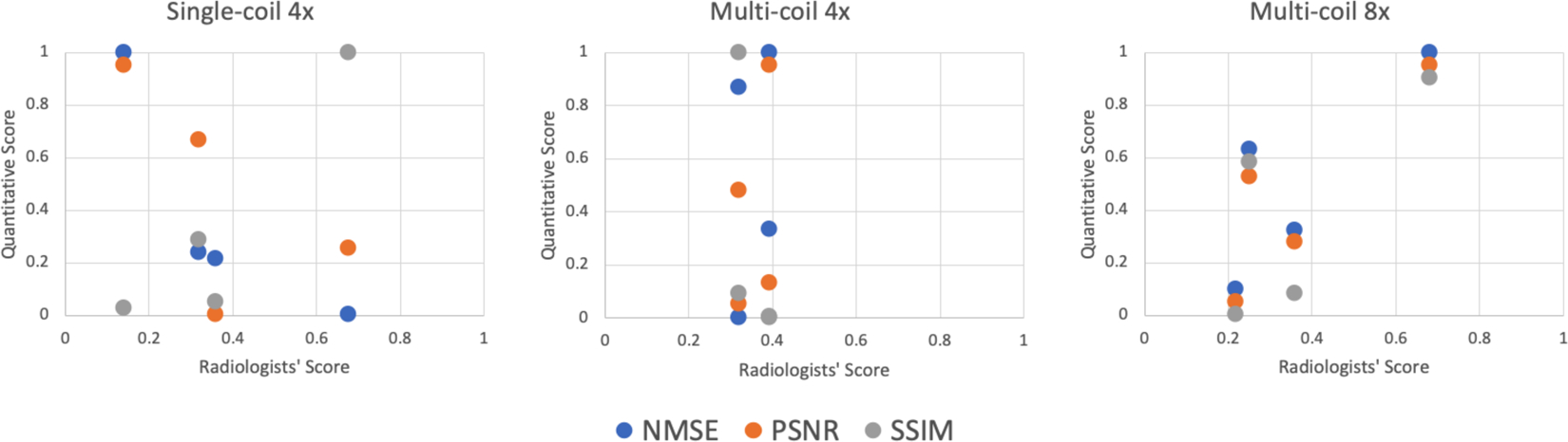
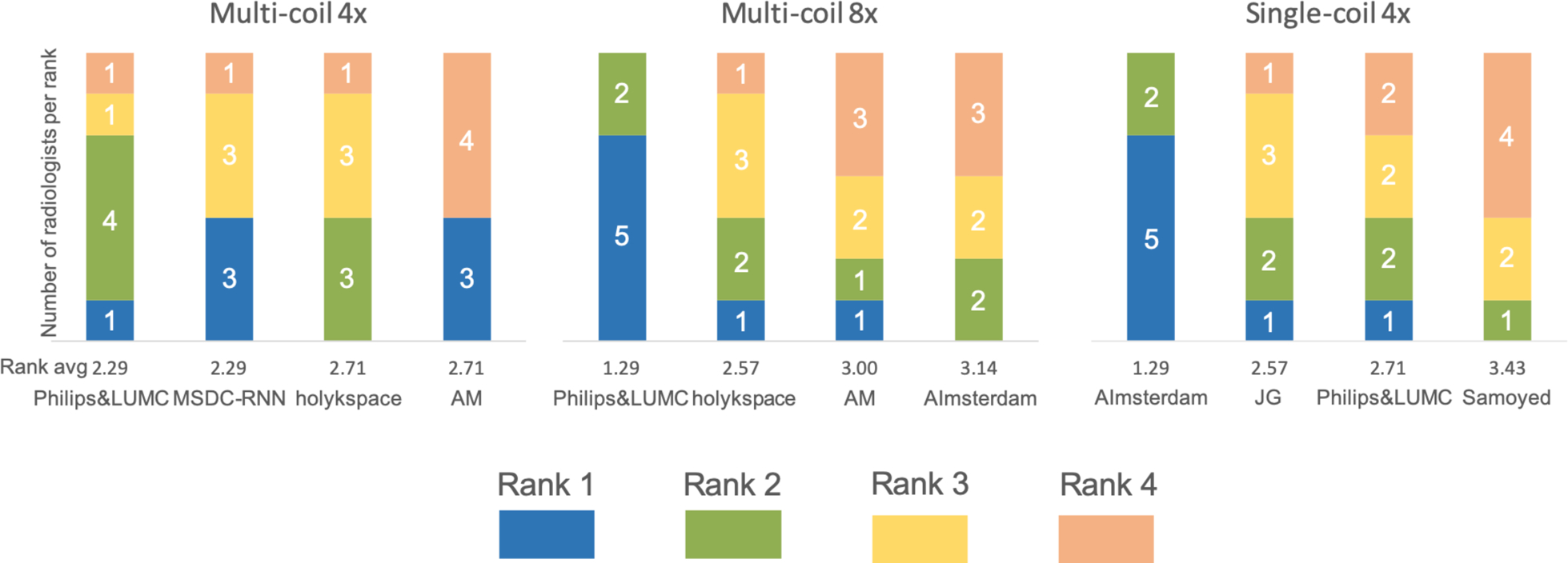
Methods: We provided participants with a dataset of raw k-space data from 1,594 consecutive clinical exams of the knee. The goal of the challenge was to reconstruct images from these data. In order to strike a balance between realistic data and a shallow learning curve for those not already familiar with MR image reconstruction, we ran multiple tracks for multi-coil and single-coil data. We performed a two-stage evaluation based on quantitative image metrics followed by evaluation by a panel of radiologists. The challenge ran from June to December of 2019.

Results: We received a total of 33 challenge submissions. All participants chose to submit results from supervised machine learning approaches.

Conclusions: The challenge led to new developments in machine learning for image reconstruction, provided insight into the current state of the art in the field, and highlighted remaining hurdles for clinical adoption.

Keywords: challenge; compressed sensing; fast imaging; image reconstruction; machine learning, optimization; parallel imaging; public dataset.


Figures

FIGURE 1
FIGURE 1
a) Online leaderboard at the completion of the challenge (December 2019). Three quantitative metrics are provided for R=4 and R=8 for both image contrasts on the webpage, along with a selection of reconstructed images. A short description of the reconstruction approach used, together with links to a corresponding paper or code repository, are also shown if the submitting groups provide this information. b) SSIM scores of the challenge submissions for each track. As expected, there is a substantial difference in overall SSIM values between the multi-coil and the single-coil tracks.
FIGURE 2
FIGURE 2
Multi-Coil R=4 track results: Selected results from the top 4 submissions in each track, for both image contrasts. The submissions are ordered from left to right based on the average of radiologists’ rankings. A combined parallel imaging and compressed sensing reconstruction using Total Generalized Variation (PI-CS) is shown for reference. SSIM to the ground truth for this particular slice is displayed in the bottom-left corner of each image. First row: Results for one slice from an acquisition without fat suppression. This case shows subtle pathology in the ROI indicated by a white rectangle in the ground truth image. Second row: One slice from an acquisition with fat suppression. Third row: Zoomed view of the ROI that shows a subchondral osteophyte (highlighted by a white arrow in the ground truth reconstruction). This pathology is not visible in any of the accelerated reconstructions.
FIGURE 3
FIGURE 3
Multi-Coil R=8 track results: Selected results from the top 4 submissions in each track. The submissions are ordered from left to right based on the average of radiologists’ rankings. A combined parallel imaging and compressed sensing reconstruction using Total Generalized Variation (PI-CS) is shown for reference. SSIM to the ground truth for this particular slice is displayed in the bottom-left corner of each image. First row: Results for one slice from an acquisition without fat suppression. This case shows shows moderate artifact from a metal implant. Second row: One slice from an acquisition with fat suppression. This case shows shows a meniscal tear in the ROI indicated by a white rectangle in the ground truth image. Third row: Zoomed view of the ROI that shows a meniscal tear (highlighted by a white arrow in the ground truth reconstruction). This pathology is not well seen in any of the accelerated reconstructions.
FIGURE 4
FIGURE 4
Single-Coil R=4 track results: Selected results from the top 4 submissions for each track. SSIM to the ground truth for this particular slice is displayed in the bottom-left corner of each image. A combined parallel imaging and compressed sensing reconstruction using Total Generalized Variation (PI-CS) is shown for reference.
FIGURE 5
FIGURE 5
Results from the case with the lowest averaged SSIM over the whole image volume from the top 4 submissions for each track. A combined parallel imaging and compressed sensing reconstruction using Total Generalized Variation (PI-CS) is shown for reference. Notably, all methods performed worst on the same case within each track, and all methods outperformed the PI-CS reference. SSIM to the ground truth for the shown slice is displayed in the bottom-left corner of each image.
FIGURE 6
FIGURE 6
Scatterplots of the quantitative scores vs the average radiologists’ score based on ranking (1 is best) for the top 4 submissions in all three submission tracks. NMSE, PSNR, and SSIM scores are normalized so that the best score corresponds to a value of 1 for convenient visualization. For multi-coil R=8, the highest ranked submission was also the one that had the highest SSIM, NMSE and PSNR values. For single-coil R=4, only SSIM showed a similar trend as the radiologists scores, while the other two metrics showed almost opposite trends. For the multi-coil R=4 track, the top 4 submissions were very close together with all metrics. Therefore, the results are less conclusive.
FIGURE 7
FIGURE 7
Individual rankings by the 7 radiologists for the top 4 submissions in all three submission tracks. The ranks from 1 to 4 are color coded, and the number of radiologists who scored each submission at a certain rank is shown in the plot. For multi-coil R=8 and single-coil R=4 tracks, the radiologists had a strong preference for a single submission. The results are less consistent for the multi-coil R=4 track.

References

    1. Kang E, Min J, Ye JC. A deep convolutional neural network using directional wavelets for low-dose X-ray CT reconstruction. Medical Physics 2017;44(10):e360–e375. - PubMed
    1. Chen H, Zhang Y, Kalra MK, Lin F, Chen Y, Liao P, et al. Low-Dose CT with a residual encoder-decoder convolutional neural network. IEEE Transactions on Medical Imaging 2017;. - PMC - PubMed
    1. Jin KH, McCann MT, Froustey E, Unser M. Deep convolutional neural network for inverse problems in imaging. IEEE Transactions on Image Processing 2017;. - PubMed
    1. Wolterink JM, Leiner T, Viergever MA, Isgum I. Generative Adversarial Networks for Noise Reduction in Low-Dose CT. IEEE transactions on medical imaging 2017;36(12):2536–2545. - PubMed
    1. Kobler E, Klatzer T, Hammernik K, Pock T. Variational Networks: Connecting Variational Methods and Deep Learning. In: Proceedings of the German Conference on Pattern Recognition (GCPR); 2017. p. 281–293.

Publication types