Review
. 2020 Jun;145(6):1485-1497.
doi: 10.1016/j.jaci.2020.02.021.

Cutaneous barrier dysfunction in allergic diseases

Affiliations
Review

Cutaneous barrier dysfunction in allergic diseases

Donald Y M Leung et al. J Allergy Clin Immunol. 2020 Jun.

Erratum in

  • Corrigenda.
    [No authors listed] [No authors listed] J Allergy Clin Immunol. 2021 Sep;148(3):905. doi: 10.1016/j.jaci.2021.06.014. J Allergy Clin Immunol. 2021. PMID: 34489014 No abstract available.

Abstract

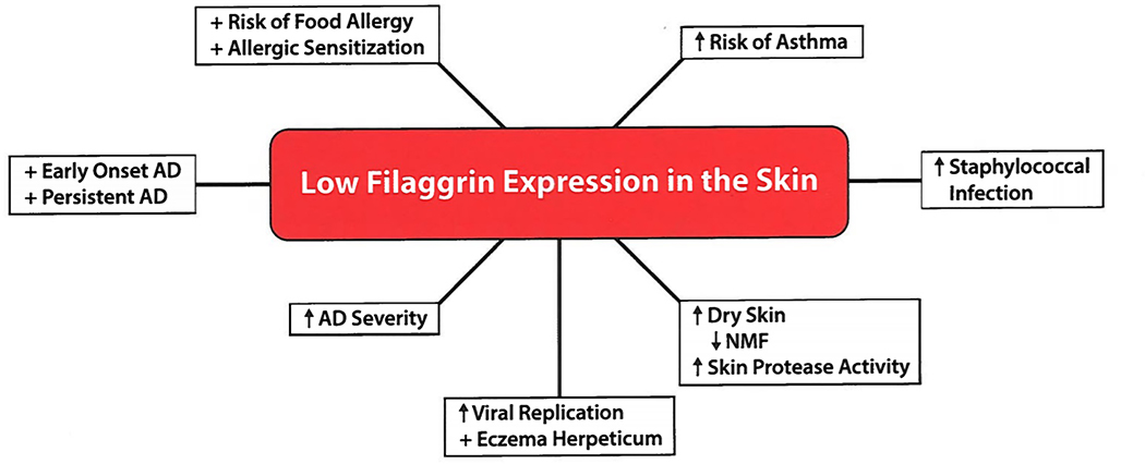
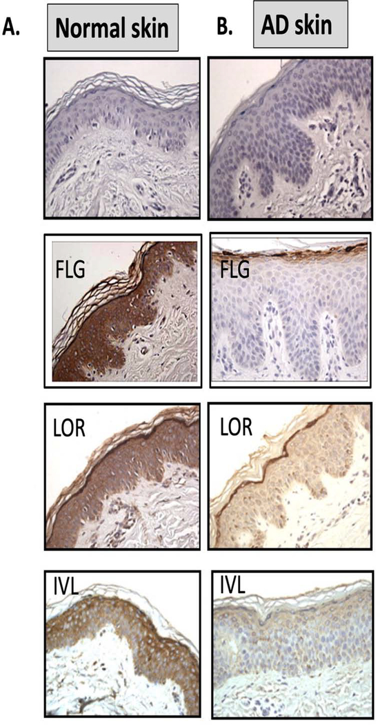
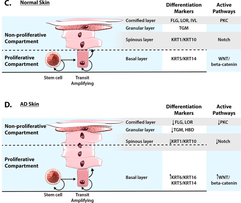
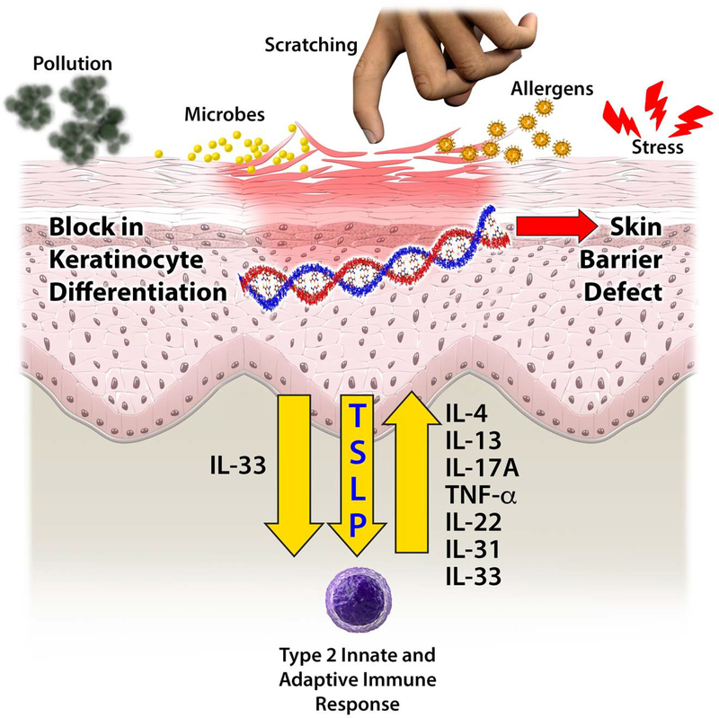
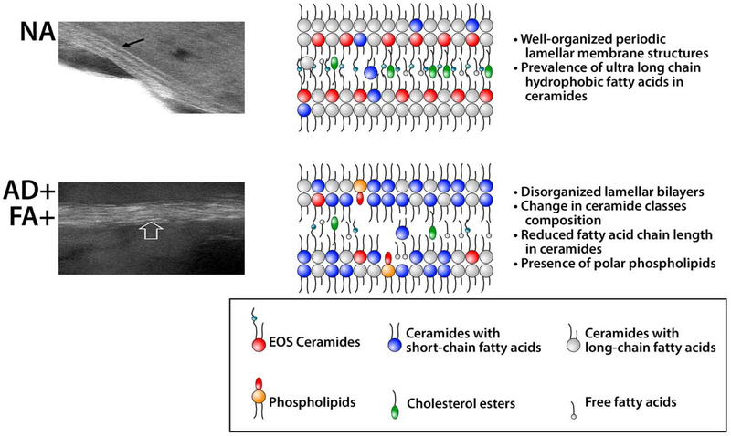
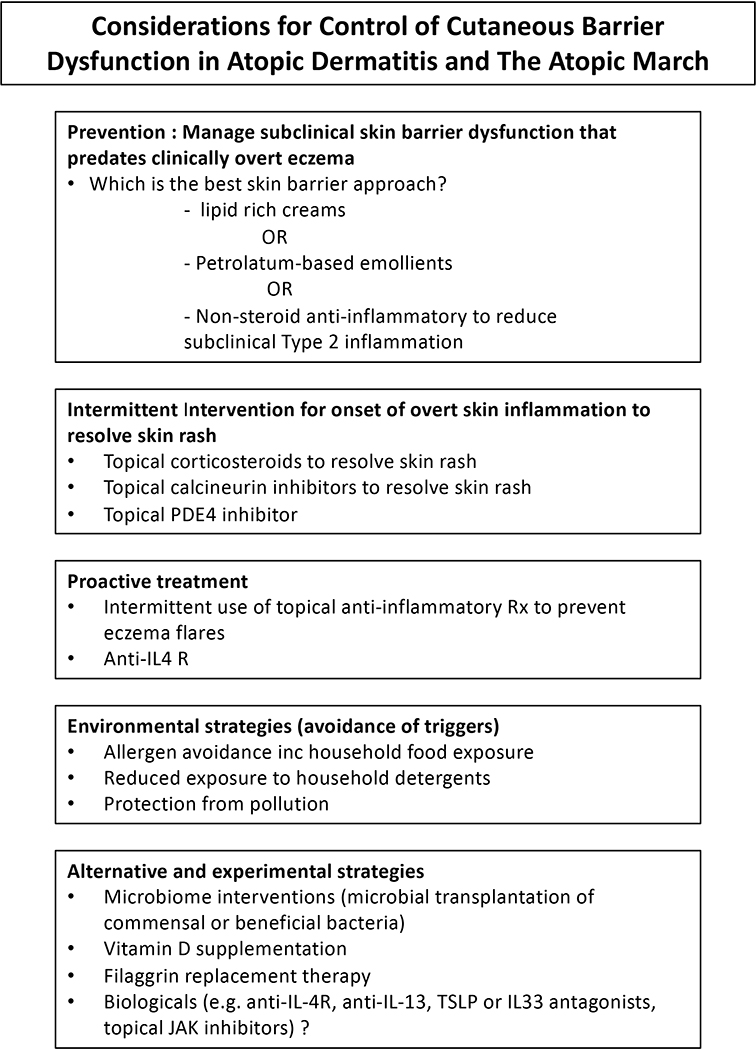
The fundamental defect(s) that drives atopic dermatitis (AD) remains controversial. "Outside in" proponents point to the important association of filaggrin gene mutations and other skin barrier defects with AD. The "inside out" proponents derive support from evidence that AD occurs in genetic animal models with overexpression of type 2 immune pathways in their skin, and humans with gain-of-function mutations in their type 2 response develop severe AD. The observation that therapeutic biologics, targeting type 2 immune responses, can reverse AD provides compelling support for the importance of "inside out" mechanisms of AD. In this review, we propose a central role for epithelial cell dysfunction that accounts for the dual role of skin barrier defects and immune pathway activation in AD. The complexity of AD has its roots in the dysfunction of the epithelial barrier that allows the penetration of allergens, irritants, and microbes into a cutaneous milieu that facilitates the induction of type 2 immune responses. The AD phenotypes and endotypes that result in chronic skin inflammation and barrier dysfunction are modified by genes, innate/adaptive immune responses, and different environmental factors that cause skin barrier dysfunction. There is also compelling evidence that skin barrier dysfunction can alter the course of childhood asthma, food allergy, and allergic rhinosinusitis. Effective management of AD requires a multipronged approach, not only restoring cutaneous barrier function, microbial flora, and immune homeostasis but also enhancing skin epithelial differentiation.

Keywords: Atopic dermatitis; epithelial barrier; food allergy; peanut allergy; skin barrier.


Figures

Figure 1.
Figure 1.
Consequences of Filaggrin Deficiency on Atopic Dermatitis and Modifying the Course of Allergic Diseases.
Figure 2.
Figure 2.. Lack of Keratinocyte Terminal Differentiation in Atopic Dermatitis.
Abnormalities in keratinocyte differentiation in AD skin result in hyperplasia of the basal layer, reduction of spinous layer and inhibition of markers of terminal differentiation (FLG, IVL, LOR) in AD (B) as compared to NA skin (A) (reproduction with permission from reference and reference 134). Schematic of epidermal differentiation pattern in normal (C) and AD skin (D) is shown. The keratinocyte differentiation process is an integrated multi-step program of sequential changes in gene expression and cell structure, as the cells migrate from the proliferative basal layer, through spinous and granular layers, into the cornified layer, which functions as a skin barrier. Cells proliferate in the basal layer of epidermis. In the spinous (suprabasal) layer, cells irreversibly exit the cell cycle and switch from KRT5/KRT14 to KRT1/KRT10 production. Wnt/beta-catenin pathway is active in the proliferating epidermis, while keratinocyte differentiation in the spinous layer is under control of the Notch pathway. Deficient Notch activity alters epidermal differentiation in AD skin, expanding the proliferative compartment and influencing subsequent changes in epidermal differentiation program. Changes in extracellular Ca2+ and lipid metabolism trigger the protein kinase C (PKC) pathway activation and regulates transcription of late differentiation markers in granular layer, FLG, LOR, IVL, HBD and TGM1. Inhibition of terminal differention marker expression is observed in AD skin. Permissions obtained from reference and reference to publish edited portions of images.
Figure 3.
Figure 3.. Causes of Cutaneous Barrier Dysfunction in AD and Allergic Diseases.
This includes genetic mutations, environmental influences (including microbes, scratching allergens) and the immune response (including IL4, IL13, IL17A, TNF-α, IL22, TSLP, IL31, IL33).
Figure 4.
Figure 4.. Lipid Barrier Abnormalities in AD Skin.
The intercellular lipids (the “mortar”) are an integral component of the stratum corneum skin barrier. They consist of a heterogeneous mixture of ceramides, free fatty acids, and cholesterol. These lipids are produced in the stratum granulosum and stored in lamellar bodies, and then secreted into extracellular space in the transition to the stratum corneum. In healthy skin nonatopic (NA), lipid lamellae are well organized; EOS Ceramides and ceramides with ultra long-chain fatty acids form a crystalline structure. In AD skin, the increase in ceramides with short-chain fatty acids as well as the presence of polar lipids disrupt lamella structure. Gaps in lipid lamellae structures of AD skin may support penetration of allergens and water loss through skin barrier. Electron microscopy photographs of NA and AD skin are reproduced from previously published work (120). Permission obtained from reference to publish edited portions of images.
Figure 5.
Figure 5.
Management Strategies for the Prevention and Treatment of AD and The Atopic March

References

    1. Martin PE, Koplin JJ, Eckert JK, Lowe AJ, Ponsonby AL, Osborne NJ, et al. The prevalence and socio-demographic risk factors of clinical eczema in infancy: a population-based observational study. Clin Exp Allergy 2013;43(6):642–51. DOI: 10.1111/cea.12092. - DOI - PubMed
    1. Holgate ST, Wenzel S, Postma DS, Weiss ST, Renz H, and Sly PD. Asthma. Nat Rev Dis Primers 2015;1:15025 DOI: 10.1038/nrdp.2015.25. - DOI - PMC - PubMed
    1. Peters RL, Koplin JJ, Gurrin LC, Dharmage SC, Wake M, Ponsonby AL, et al. The prevalence of food allergy and other allergic diseases in early childhood in a population-based study: HealthNuts age 4-year follow-up. J Allergy Clin Immunol 2017;140:145–53. DOI: 10.1016/j.jaci.2017.02.019. - DOI - PubMed
    1. Walker MT, Green JE, Ferrie RP, Queener AM, Kaplan MH, Cook-Mills JM. Mechanism for initiation of food allergy: dependence on skin barrier mutations and environmental allergen costimulation. J Allergy Clin Immunol 2018;141(5):1711–25. DOI: 10.1016/j.jaci.2018.02.003. - DOI - PMC - PubMed
    1. Kennedy K, Heimall J, Spergel JM. Advances in atopic dermatitis in 2017. J Allergy Clin Immunol 2018;142(6):1740–7. DOI: 10.1016/j.jaci.2018.10.012. - DOI - PubMed

Publication types