

Coats-like Exudative Vitreoretinopathy in Retinitis Pigmentosa: Ocular Manifestations and Treatment Outcomes
- PMID: 32507488
- PMCID: PMC8086515
- DOI: 10.1016/j.oret.2020.03.026
Coats-like Exudative Vitreoretinopathy in Retinitis Pigmentosa: Ocular Manifestations and Treatment Outcomes
Abstract
Purpose: To provide a comprehensive review of the ocular manifestations, outcomes, and genetic findings in patients with Coats-like retinitis pigmentosa (RP).
Design: Multicenter, retrospective, nonconsecutive case series.
Participants: Patients with a diagnosis of RP demonstrating Coats-like exudative vitreoretinopathy between January 1, 2008, and October 1, 2019.
Methods: Evaluation of ocular findings at RP diagnosis and at time of presentation of Coats-like exudative vitreoretinopathy, pedigree analysis, genetic testing, retinal imaging, and anatomic outcomes after treatment.
Main outcome measures: Visual acuity, ophthalmoscopy results, OCT results, fluorescein angiography results, and identification of genetic mutations.
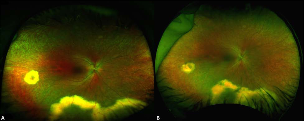
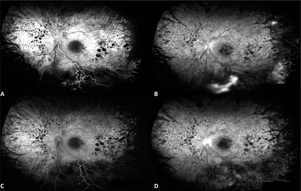
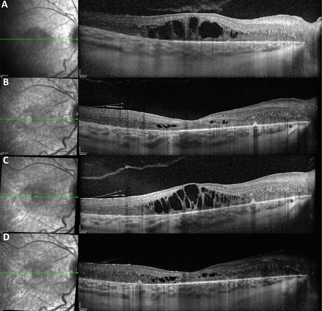
Results: Nine patients diagnosed with RP and demonstrating Coats-like exudative vitreoretinopathy were included. Median age at time of RP diagnosis was 8 years (range, 1-22 years), and median age at presentation of Coats-like exudative vitreoretinopathy was 18 years (range, 1-41 years). Seven patients were female, and 2 were male. The genetic cause of disease was identified in 6 patients. Three patients demonstrated Coats-like fundus findings at the time of RP diagnosis. Exudative retinal detachment (ERD) localized to the infratemporal periphery was present in all patients, with bilateral disease observed in 7 patients. In all treated patients, focal laser photocoagulation was used to treat leaking telangiectasias and to limit further ERD expansion. Cystoid macular edema refractory to carbonic anhydrase inhibitor therapy and ultimately amenable to treatment with intravitreal anti-vascular endothelial growth factor injection was observed in 4 patients.
Conclusions: Coats-like vitreoretinopathy is present in up to 5% of all RP patients. The term Coats-like RP is used colloquially to describe this disease state, which can present at the time of RP diagnosis or, more commonly, develops late during the clinical course of patients with longstanding RP. Coats-like RP is distinct from Coats disease in that exudative pathologic features occur exclusively in the setting of a coexisting RP diagnosis, is restricted to the infratemporal retina, can affect both eyes, and does not demonstrate a male gender bias. Given the risk of added vision loss posed by exudative vitreoretinopathy in patients with RP, a heightened awareness of this condition is critical in facilitating timely intervention.
Copyright © 2020 American Academy of Ophthalmology. Published by Elsevier Inc. All rights reserved.
Conflict of interest statement
Figures
References
-
- Weleber RG, Gregory-Evans K. Retinitis Pigmentosa and Allied Disorders. Retina. 2006:395–498.
-
- Zamorani G. Una rara associazone di retinite di Coats con retinite pigmentosa. G Ital Oftalmol. 1956;9:429–443.
-
- Khan JA, Ide CH, Strickland MP. Coats’-type retinitis pigmentosa. Surv Ophthalmol. 1988;32:317–332. - PubMed
-
- Egerer I, Tasman W, Tomer TT. Coats disease. Arch Ophthalmol. 1974;92:109–112. - PubMed
Publication types
MeSH terms
Grants and funding
LinkOut - more resources
Full Text Sources
Other Literature Sources
Medical
