Review
. 2020 Aug;28(8):605-618.
doi: 10.1016/j.tim.2020.05.012. Epub 2020 May 20.

Spiking Pandemic Potential: Structural and Immunological Aspects of SARS-CoV-2

Affiliations
Review

Spiking Pandemic Potential: Structural and Immunological Aspects of SARS-CoV-2

Ying-Ting Wang et al. Trends Microbiol. 2020 Aug.

Abstract

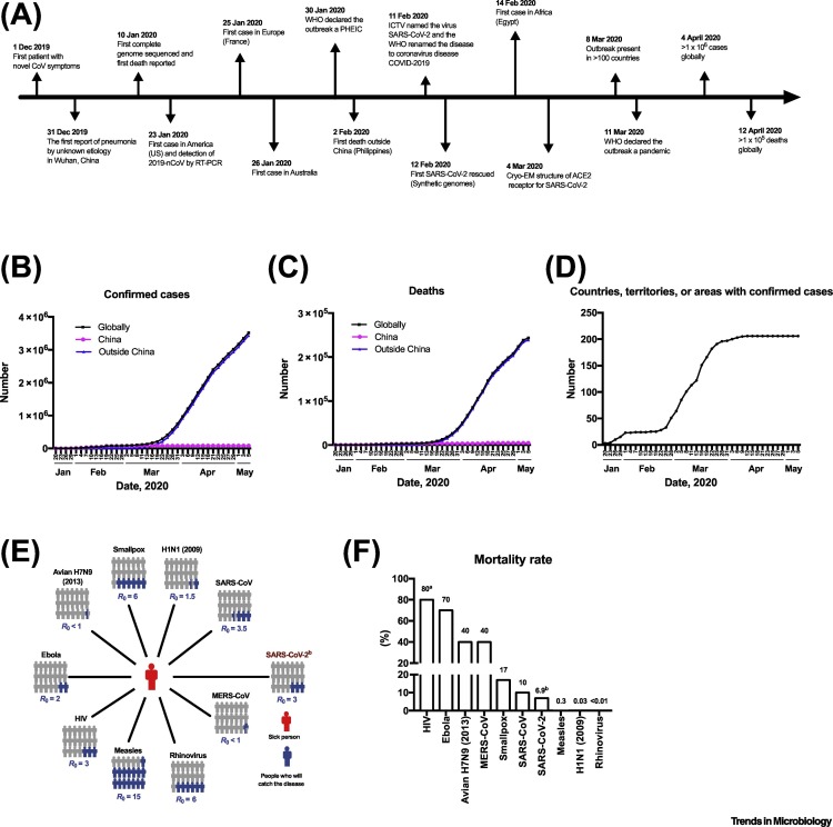
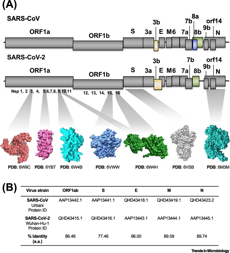
SARS-Coronavirus-2 (SARS-CoV-2) causes Coronavirus disease 2019 (COVID-19), an infectious respiratory disease causing thousands of deaths and overwhelming public health systems. The international spread of SARS-CoV-2 is associated with the ease of global travel, and societal dynamics, immunologic naiveté of the host population, and muted innate immune responses. Based on these factors and the expanding geographic scale of the disease, the World Health Organization (WHO) declared the COVID-19 outbreak a pandemic-the first caused by a coronavirus. In this review, we summarize the current epidemiological status of COVID-19 and consider the virological and immunological lessons, animal models, and tools developed in response to prior SARS-CoV and MERS-CoV outbreaks that can serve as resources for development of SARS-CoV-2 therapeutics and vaccines. In particular, we discuss structural insights into the SARS-CoV-2 spike protein, a major determinant of transmissibility, and discuss key molecular aspects that will aid in understanding and fighting this new global threat.

Keywords: COVID-19; SARS-CoV-2; coronavirus; host immune response; pandemic; spike.


Figures

Figure 1
Figure 1
Timeline of Major Events for Severe Acute Respiratory Syndrome-Coronavirus-2 (SARS-CoV-2), Total Numbers of Coronavirus Disease 2019 (COVID-19) Cases, and Basic Reproduction Number (R0) and Mortality Rate of Select Viruses. (A) Timeline of events, (B) cumulative confirmed cases, (C) reported deaths, and (D) countries with reported cases. Squares indicates the total number of COVID-19 cases reported worldwide. Circles and triangles indicate the number of COVID-19 cases reported in China and in other countries, respectively. Data from WHO as of May 5 2020. (E) R0 value indicates the number of people (colored in blue) infected from one contagious person (colored in red). (F) Case fatality rate (CFR) of select viruses. aCFR is based on patients not receiving therapy. bData on SARS-CoV-2 are derived from WHO. Abbreviations: Cryo-EM, Cryogenic electron microscopy; ICTV, International Committee on Taxonomy of Viruses; PHEIC, public health emergency of international concern; WHO, World Health Organization.
Figure 2
Figure 2
Genomic Distribution of Severe Acute Respiratory Syndrome-Coronavirus-2 (SARS-CoV-2) and SARS-CoV. (A) The genomes of SARS-CoV (upper panel) and SARS-CoV-2 (lower panel) are shown as lines and the open reading frames (ORFs) are represented by gray and colored boxes to indicate those that have similar and different lengths, respectively. The atomic structure for some of the SARS-CoV-2 proteins is shown in surface representation; main protease 3CLpro or nonstructural protein 5 (nsp5) with unliganded active site in pink, nsp9 RNA binding protein in cyan, nsp15 endoribonuclease in marine blue, nsp16–nsp10 complex in green, prefusion spike glycoprotein in gray, and nucleocapsid protein N terminal RNA binding domain in deep turquoise. For visual clarity, the length of the boxes is not proportional to the real sequence length and the atomic structures are not proportional to their molecular weight. (B) Percentage identity matrix for the alignment of SARS-CoV and SARS-CoV-2 amino acids. Abbreviations: PDB, Protein Data Bank.
Figure 3
Figure 3
Coronavirus (CoV) Life Cycle and Host Immune Response. The CoV life cycle initiates with the binding of spike proteins on the virion surface to their specific receptor [e.g., ACE2 for severe acute respiratory syndrome-coronavirus-2 (SARS-CoV-2)] on target cells. Following receptor binding and endocytosis, S protein undergoes a conformational change and viral genomic RNA is released into the cytoplasm. Viral genomic RNA functions as mRNA, which is translated into polyproteins (pp) pp1a and pp1ab, which are then proteolytically cleaved into mature nonstructural proteins (nsps). Many nsps synergize to modify the endoplasmic reticulum (ER) membrane to form double membrane vesicles (DMVs) where transcription of genomic and subgenomic RNA occurs. The truncated subgenomic RNA of the 5′-ends are used as templates to translate structural (S, E, M, and N) and accessory proteins. S, E, and M assemble together with nucleocapsid (one copy of viral genome encapsulated by N proteins) at the ER–Golgi intermediate compartment (ERGIC) and viral progeny are released by exocytosis. Host immune responses are triggered by danger signals from infected cells or free virions, which are recognized by innate immune cells. Elevated proinflammatory cytokines and chemokines can be seen in asymptomatic to mild cases. Exuberated cytokine production resulting in cytokine storm exacerbates the severity of coronavirus disease 2019 (COVID-19). Lymphopenia (T and B cells) and neutrophil infiltration into infected sites contribute to the pathogenesis of COVID-19. Several CoV proteins have been reported to be capable of inhibiting the type I IFN signaling pathway. Figure created with BioRender (biorender.com).
Figure 4
Figure 4
Atomic Structure of Severe Acute Respiratory Syndrome-Coronavirus-2 (SARS-CoV-2) Receptor-Binding Domain (RBD). (A) Schematic architecture of the SARS-CoV-2 spike glycoprotein. Degree of protein surface conservation between trimeric SARS-CoV-2 and SARS-CoV spike protein. A color range is shown with green and magenta representing not conserved and highly conserved, respectively. The atomic position of the cleavage site is indicated by an arrow. (B) Cartoon representation of a structural alignment of the SARS-CoV (gray) and SARS-CoV-2 (green) RBDs interacting with ACE2 receptor (orange) shown in surface representation. Corresponding footprints of SARS-CoV RBD and SARS-CoV-2 RBD overlaid on the ACE2 receptor are colored in gray and green, respectively, to illustrate overlap between both interaction sites. A differential loop between SARS-CoV-2 and SARS-CoV is indicated with a dashed circle. (C) Closed and opened conformation of the SARS-CoV-2 S with one of the RBD domains buried or exposed, respectively (yellow). Abbreviations: FP, Fusion peptide; HR, heptad repeat regions; RRAR, unique furin cleavage site; SP, signal peptide; TM, transmembrane domain.

References

    1. Gorbalenya A.E. The species severe acute respiratory syndrome-related coronavirus: classifying 2019-nCoV and naming it SARS-CoV-2. Nat. Microbiol. 2020;5:536–544. - PMC - PubMed
    1. Chan J.F. A familial cluster of pneumonia associated with the 2019 novel coronavirus indicating person-to-person transmission: a study of a family cluster. Lancet. 2020;395:514–523. - PMC - PubMed
    1. Heffernan J.M. Perspectives on the basic reproductive ratio. J. R. Soc. Interface. 2005;2:281–293. - PMC - PubMed
    1. Chen J. Pathogenicity and transmissibility of 2019-nCoV-A quick overview and comparison with other emerging viruses. Microbes Infect. 2020;22:69–71. - PMC - PubMed
    1. Bai Y. Presumed asymptomatic carrier transmission of COVID-19. JAMA. 2020 doi: 10.1001/jama.2020.2565. Published online February 21, 2020. - DOI - PMC - PubMed

Publication types

MeSH terms

Substances