. 2020 Jan;10(1):57-73.
doi: 10.1002/ctm2.19. Epub 2020 Apr 7.

ATPase copper transporter A, negatively regulated by miR-148a-3p, contributes to cisplatin resistance in breast cancer cells

Affiliations

ATPase copper transporter A, negatively regulated by miR-148a-3p, contributes to cisplatin resistance in breast cancer cells

Ze Yu et al. Clin Transl Med. 2020 Jan.

Abstract

Background: Breast cancer is the leading cause of death among women. Cisplatin is an effective drug for breast cancer, but resistance often develops during long term chemotherapy. While the mechanism of chemotherapy resistance is still not fully understood.

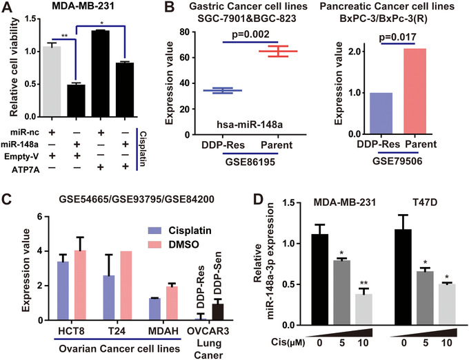
Methods: Survival analyses of ATP7A and ATP7B were used to evaluate their effects on the development of Breast invasive carcinoma (BRCA). Immunostaining, flow cytometry, and IC50 assay were utilized to examine the effects of ATP7A-siRNA combined with cisplatin on apoptosis in breast cancer cells. Q-PCR, western blotting, and dual-luciferase assay were employed to confirm ATP7A is a novel target gene of miR-148a-3p.

Results: In this current study, we identified knocking-down ATP7A could enhance cytotoxicity treatment of cisplatin in breast cancer cells. We also demonstrated miR-148a-3p overexpression in BRCA cells increased the sensitivity to cisplatin, and subsequently enhanced DNA damage and apoptosis. Moreover, we found ATP7A is a novel target gene of miR-148a-3p. In brief, our results showed miR-148a could accelerate chemotherapy induced-apoptosis in breast cancer cells by inhibiting ATP7A expression.

Conclusions: Our results highlight that inhibition of ATP7A is a potential strategy for targeting breast cancer resistant to cisplatin, and we provided an interesting method to compare the involvement of various genes in the assessment of cisplatin resistance.

Keywords: ATP7A; breast cancer; chemoresistance; cisplatin; miR-148a-3p.


Conflict of interest statement

The authors declare that there is no conflict of interest that could be perceived as prejudicing the impartiality of the research reported.

Figures

FIGURE 1
FIGURE 1
ATP7A is a potential oncogene in breast cancer. A, The schematic model of ATP7A/ATP7B regulation of cancer cell cisplatin‐resistance. B, Pearson correlation analysis between mRNA levels of ATP7A and ATP7B in BRCA samples (data came from ENCORI, cBioPortal, LinkedOmics and GEPIA). C, Analysis of ATP7A and ATP7B expression in BRCA and corresponding normal tissues using GEPIA and linkedOmics database. D, Survival curves analysis for breast cancer patients with low versus high expression levels of ATP7A and ATP7B. E, Heat map shows the expression of ATP7A and ATP7B along samples in risk groups. Low expression is represented in green grades and high expression in red grades; box plots shows the expression of ATP7A and ATP7B genes in risk groups, including the P‐value testing for difference using t‐test. F, Enrichment analysis of ATP7A and ATP7B related genes, respectively
FIGURE 2
FIGURE 2
ATP7A induces cisplatin resistance in breast cancer cells. A, MDA‐MB‐231 cells were transfected with ATP7A or ATP7B siRNAs, and ATP7A or ATP7B expression was examined by western blot. B, Cell viability was determined using CCK‐8 in T47D and MDA‐MB‐231 cells transfected with si‐ATP7As or si‐ATP7Bs. C, Flow cytometry analysis of apoptosis rates in MDA‐MB‐231 (with si‐ATP7As) cells with or without cisplatin treatment. D, Cisplatin induces morphology changes in MDA‐MB‐231 cells transfected with ATP7A‐siRNAs. E, Cisplatin IC50s calculated using CCK‐8 in breast cancer cells transfected with si‐ATP7As or si‐nc. F, Western blot was used to detect overexpression of ATP7A. G, Cisplatin IC50s calculated using CCK‐8 in cells treated with overexpressed ATP7A. H, Immunofluorescence of P‐H2AX was used to evaluate the degree of DNA‐damage in cisplatin‐treated MDA‐MB‐231 cells transfected with ATP7A‐siRNAs. Each bar in the figure represents the mean ± SEM of triplicates. ** P < .01
FIGURE 3
FIGURE 3
Microarray analysis of TNBC cells treated with cisplatin. A, The schematic model of knocked‐down ATP7A increased cancer cell sensitivity to cisplatin. B, MCLP database showed Protein‐drug (cisplatin) correlation analysis in BRCA. C and D, RT‐PCR analysis of ATM, BID, HSP70, Bcl2 and BclA1 expression levels in cisplatin‐treated MDA‐MB‐231 cells transfected with ATP7A‐siRNAs, compared with the cells treated with cisplatin alone. E, Bubble chart showing the top enriched pathways by DAVID enrichment analysis (functional categories) to demonstrate the difference in gene enrichment in cisplatin‐induced TNBC cells. Each bar in the figure represents the mean ± SEM of triplicates. * P < .05, ** P < .01, and *** P < .001
FIGURE 4
FIGURE 4
Cisplatin does not affect intracellular copper levels. A, The methylation level of ATP7A in BRCA samples is shown (Column 1 represents BRCA samples, Column 2 represents ATP7A expression, Column 3 represents the DNA methylation clusters in corresponding sample). B, The schematic model of main pathways of intracellular copper metabolism. C, The mRNA expression levels of SLC31A1, Atox1, CCS, Cox17 were decreased in MDA‐MB‐231 cells treated with CuSO4, while ATP7A and ATP7B increased with the same condition. Accordingly, TM led to upregulation of SLC31A1, Atox1, CCS, Cox17 and decrease in ATP7A and ATP7B. D, The mRNA expression levels of SLC31A1, Atox1 and ATP7A were increased in MDA‐MB‐231 cells treated with cisplatin, while CCS and Cox17 showed no significant difference. E, Flow cytometry analysis was performed to detect copper level in cisplatin‐treated MDA‐MB‐231 cells, CuSO4 treatment as a positive control, while TM treatment as a negative control. Each bar in the figure represents the mean ± SEM of triplicates. * P < .05, ** P < .01, and *** P < .001
FIGURE 5
FIGURE 5
ATP7A is the target of miR‐148a‐3p. A, Venn diagram of putative miRNAs targeting ATP7A. B, Pearson correlation analysis between ATP7A and miR‐148a‐3p in BRCA samples. C and D, Expression analysis of ATP7A and miR‐148a‐3p in the same BRCA samples and cell lines. E, Survival curves analysis for breast cancer patients with high versus low expression levels of miR‐148a‐3p or miR‐148b‐4p. F, Overlap of negative correlation genes and proteins of miR‐148a‐3p. G, After treatment with doses of miR‐148a‐3p mimic for 72 hours, western blot analysis was performed to detect ATP7A protein levels in MDA‐MB‐231 cells. H, Wild‐type ATP7A luciferase reporter vector and mutant‐ATP7A luciferase reporter vector containing an 8 bp mutation in the predicted miR‐148a‐3p binding sites were constructed. I, The relative luciferase activity was significantly reduced in the 293T cells transfected with miR‐148a‐3p and these effects could be abolished by mutation of ATP7A 3'‐UTR. Each bar in the figure represents the mean ± SEM of triplicates. * P < .05, ** P < .01
FIGURE 6
FIGURE 6
The role of miR‐148a‐3p in breast cancer cell cisplatin‐resistance. A and B, cell viability and invasion assay of MDA‐MB‐231 cells treated with miR‐148a‐3p mimics. C, DAPI nuclear staining and nucleus counting were applied to detect apoptosis and cell proliferation in MDA‐MB‐231 cells transfected with miR‐148a‐3p or miR‐nc. D, Immunofluorescence staining of caspase‐3 in cisplatin‐treated MDA‐MB‐231 cells transfected with miR‐148a‐3p. E, Xenograft tumors of sacrificed mice at end of experiment with or without cisplatin treatment. F, Immunofluorescence staining of p‐H2AX in cisplatin‐treated MDA‐MB‐231 cells transfected with miR‐148a‐3p. Each bar in the figure represents the mean ± SEM of triplicates. * P < .05, ** P < .01, and *** P < .001
FIGURE 7
FIGURE 7
MiR‐148a‐3p restrains cisplatin‐resistance through the regulation of ATP7A in breast cancer cells. A, MDA‐MB‐231 cells were co‐transfected with miR‐148a‐3p or miR‐nc and empty or ATP7A‐overexpressed vectors; cell viability was determined using CCK‐8 assays. B, The expression level of miR‐148a‐3p in cisplatin‐resistant cell lines, compared with normal parent cells. C, The expression of miR‐148a‐3p in cisplatin‐treated cells, compared with cells treated with DMSO. D, Expression of miR‐148a‐3p was determined in MDA‐MB‐231 and T47D cells with the increasing doses of cisplatin for 48 hours. Each bar in the figure represents the mean ± SEM of triplicates. * P < .05, ** P < .01
FIGURE 8
FIGURE 8
ATP7A is an important target of miR‐148a. A, Expression levels of miR‐148a‐3p targets in MDA‐MB‐231 cells transfected with miR‐148a‐3p mimic were determined by RT‐PCR. B, PCR was used to validate mRNA levels altered through overexpression of the corresponding siRNAs. C, Caspase‐3 activity analysis of MDA‐MB‐231 cells treated with cisplatin and multiple siRNAs, respectively, by western blot. D, Caspase‐3 activated probe was used to detect apoptosis in MDA‐MB‐231 cells treated with cisplatin and multiple siRNAs, respectively
FIGURE 9
FIGURE 9
Schematic diagram of the mechanism that ATP7A is involved in cisplatin resistance. The blue arrow represents the efflux of cisplatin through MRPs; the red arrow represents the efflux of cisplatin through ATP7A in our present study

References

    1. Anastasiadi Z, Lianos GD, Ignatiadou E, Harissis HV, Mitsis M. Breast cancer in young women: an overview. Updates Surg. 2017;69:313‐317. - PubMed
    1. Colditz GA, Bohlke K. Priorities for the primary prevention of breast cancer. CA Cancer J Clin. 2014;64:186‐194. - PubMed
    1. Majidinia M, Yousefi B. DNA repair and damage pathways in breast cancer development and therapy. DNA Repair (Amst). 2017;54:22‐29. - PubMed
    1. Weitzel JN. The genetics of breast cancer: what the surgical oncologist needs to know. Surg Oncol Clin N Am. 2015;24:705‐732. - PubMed
    1. Fan L, Strasser‐Weippl K, Li JJ, et al. Breast cancer in China. Lancet Oncol. 2014;15: e279‐e289. - PubMed

LinkOut - more resources