

Histopathological features and prevalence of Capillaria hepatica infection in Rattus spp. in Philippine Mount Makiling forest reserve and its adjacent areas
- PMID: 32508408
- PMCID: PMC7244692
- DOI: 10.1007/s12639-019-01189-1
Histopathological features and prevalence of Capillaria hepatica infection in Rattus spp. in Philippine Mount Makiling forest reserve and its adjacent areas
Abstract
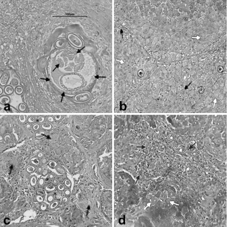
Rats are recognized as reservoir hosts of several pathogens that pose a threat to human health. Although rats are reported to be hosts of a large number of pathogens, a survey of Capillaria hepatica carried by rats in various settings such as residential, agroforestry, and agricultural areas in the Philippines has not been conducted. A total of 90 rats composed of Rattus norvegicus, Rattus tanezumi, and Rattus exulans were collected through trapping in selected residential, agroforestry, and agricultural areas in Los Baños Laguna, Philippines. The overall prevalence of C. hepatica among rats was 21.11%. Among the rat species collected, R. norvegicus showed the highest prevalence (55.56%), followed by R. exulans (14.29%), then R. tanezumi (5.36%) (differences significant at p < 0.05). Moreover, residential areas had the highest prevalence of C. hepatica infection (50%), followed by agroforestry and agricultural areas at 6.7% each (significant at p < 0.05). However, the difference in C. hepatica infection between male (11.43%; 4/35) and female (27.27%; 15/55) rats was not significant (p > 0.05). Most of the infected rats were moderately infected (68.42%), while few were lightly and severely infected (15.78% each). Lastly, the presence of C. hepatica in liver is suggestive of presence of lymphocytes, amyloid, granuloma, and the occurrence of necrosis, hypertrophy, fibrosis, and cholestasis in the liver of the host. Capillariasis could be occurring in Philippine human populations, hence there is need for screening the population with appropriate means and to create awareness of this emerging disease.
Keywords: Capillaria hepatica; Histopathology; Liver; Philippines; Prevalence; Rats.
© Indian Society for Parasitology 2020.
Conflict of interest statement
Conflict of interestThe authors declare that they have no conflict of interest.
Figures


References
-
- Aghdam MK, Karimi A, Amanati A, Ghoroubi J, Khoddami M, Shamsian BS, Far SZ. Capillaria hepatica, A case report and review of the literatures. Arch Pediatr Infect Dis. 2015 doi: 10.5812/pedinfect.19398. - DOI
-
- Aplin KP, Brown PR, Jacob J, Krebs CJ, Singleton GR. Field methods for rodent studies in Asia and the Indo-Pacific. Canberra: Australian Centre for International Agricultural Research; 2003.
-
- Baker DG, editor. Flynn’s parasites of laboratory animals. New York: Wiley; 2008.
-
- Brooks JE, Rowe FP, World Health Organization, Division of Vector Biology and Control (1987) Rodents: training and information guide. World Health Organization
-
- Centers for Disease Control and Prevention (2012) Parasite Capillariasis (also known as Capillaria infection). https://www.cdc.gov/parasites/capillaria/faqs.html. Accessed 11 July 2017
LinkOut - more resources
Full Text Sources
Miscellaneous