Dual-Energy CT Follow-Up After Stroke Thrombolysis Alters Assessment of Hemorrhagic Complications
- PMID: 32508735
- PMCID: PMC7249255
- DOI: 10.3389/fneur.2020.00357
Dual-Energy CT Follow-Up After Stroke Thrombolysis Alters Assessment of Hemorrhagic Complications
Abstract
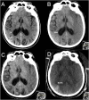
Background and Purpose: We aimed to determine whether dual-energy CT (DECT) follow-up can differentiate contrast staining (CS) from intracranial hemorrhage (ICH) in stroke patients treated with intravenous thrombolysis (IVT), who had undergone acute stroke imaging using CT angiography (CTA), and CT perfusion (CTP). Materials and Methods: Between November 2012 and January 2018, 168 patients at our comprehensive stroke center underwent DECT follow-up within 36 h after IVT and acute CTA with or without CTP but did not receive intra-arterial imaging or treatment. Two independent readers evaluated plain monochromatic CT (pCT) alone and compared this with a second reading of a combined DECT approach using pCT and water- and iodine-weighted images, establishing and grading the ICH diagnosis, per Heidelberg and Safe Implementation of Treatments in Stroke Monitoring Study (SITS-MOST) classifications. Results: On pCT alone within 36 h, 31/168 (18.5%) patients had findings diagnosed as ICH. Using combined DECT (cDECT) changed ICH diagnosis to "CS only" in 3/168 (1.8%) patients, constituting 3/31 (9.7%) of cases with initially pCT-diagnosed ICH. These three cases had pCT diagnoses of one SAH, one minor, and one more extensive petechial hemorrhage (hemorrhagic infarction types 1 and 2), respectively. pCT alone had a 100% sensitivity, 98% specificity, 90% positive predictive value (PPV), 100% negative predictive value (NPV), and 98% accuracy for any ICH, compared to the cDECT. Inter-reader agreement for ICH classification using pCT compared to DECT was weighted kappa 0.92 (95% CI 0.87-0.98) vs. 0.91 (0.85-0.95). Conclusion: Compared to pCT, DECT within 36 h after IV thrombolysis for acute ischemic stroke, changes the radiological diagnosis of post-treatment ICH to "CS only" in a small proportion of patients. Studies are warranted of whether the altered radiological reports have an impact on patient management, for example initiation timing of antithrombotic secondary prevention.
Keywords: acute ischemic stroke; computed tomography; intracerebral hemorrhage (ICH); intravenous thrombolysis; spectral computed tomography.
Copyright © 2020 Almqvist, Almqvist, Holmin and Mazya.
Figures


Similar articles
-
Dual energy CT after stroke thrombectomy alters assessment of hemorrhagic complications.Neurology. 2019 Sep 10;93(11):e1068-e1075. doi: 10.1212/WNL.0000000000008093. Epub 2019 Aug 13. Neurology. 2019. PMID: 31409735
-
A Novel Dual-Energy CT Method for Detection and Differentiation of Intracerebral Hemorrhage From Contrast Extravasation in Stroke Patients After Endovascular Thrombectomy : Feasibility and First Results.Clin Neuroradiol. 2023 Mar;33(1):171-177. doi: 10.1007/s00062-022-01198-3. Epub 2022 Aug 12. Clin Neuroradiol. 2023. PMID: 35960327 Free PMC article.
-
Initial experience with dual-layer detector spectral CT for diagnosis of blood or contrast after endovascular treatment for ischemic stroke.Neuroradiology. 2022 Jan;64(1):69-76. doi: 10.1007/s00234-021-02736-5. Epub 2021 May 27. Neuroradiology. 2022. PMID: 34046731
-
Intracerebral hemorrhage secondary to intravenous and endovascular intraarterial revascularization therapies in acute ischemic stroke: an update on risk factors, predictors, and management.Neurosurg Focus. 2012 Apr;32(4):E2. doi: 10.3171/2012.1.FOCUS11352. Neurosurg Focus. 2012. PMID: 22463112 Review.
-
[Value of modern CT-techniques in the diagnosis of acute stroke].Radiologe. 2004 Apr;44(4):380-8. doi: 10.1007/s00117-003-1003-7. Radiologe. 2004. PMID: 15103412 Review. German.
Cited by
-
Diagnostic accuracy of dual-energy computed tomography in the diagnosis of neurological complications after endovascular treatment of acute ischaemic stroke: a systematic review and meta-analysis.Br J Radiol. 2024 Jan 23;97(1153):73-92. doi: 10.1093/bjr/tqad007. Br J Radiol. 2024. PMID: 38263833 Free PMC article.
-
Type of intracranial hemorrhage after endovascular stroke treatment: association with functional outcome.J Neurointerv Surg. 2023 Oct;15(10):971-976. doi: 10.1136/jnis-2022-019474. Epub 2022 Oct 19. J Neurointerv Surg. 2023. PMID: 36261280 Free PMC article.
-
Value of dual energy CT in post resuscitation coma. Differentiating contrast retention and ischemic brain parenchyma.Radiol Case Rep. 2022 Aug 4;17(10):3722-3726. doi: 10.1016/j.radcr.2022.07.046. eCollection 2022 Oct. Radiol Case Rep. 2022. PMID: 35965920 Free PMC article.
-
Intracranial Bleeding After Reperfusion Therapy in Acute Ischemic Stroke.Front Neurol. 2021 Feb 9;11:629920. doi: 10.3389/fneur.2020.629920. eCollection 2020. Front Neurol. 2021. PMID: 33633661 Free PMC article. Review.
References
-
- Powers WJ, Rabinstein AA, Ackerson T, Adeoye OM, Bambakidis NC, Becker K, et al. . 2018 guidelines for the early management of patients with acute ischemic stroke: a guideline for healthcare professionals from the american heart association/american stroke association. Stroke. (2018) 49:e46–e110. 10.1161/STR.0000000000000158 - DOI - PubMed
-
- Tijssen MP, Hofman PA, Stadler AA, van Zwam W, de Graaf R, van Oostenbrugge RJ, et al. . The role of dual energy CT in differentiating between brain haemorrhage and contrast medium after mechanical revascularisation in acute ischaemic stroke. Eur Radiol. (2014) 24:834–40. 10.1007/s00330-013-3073-x - DOI - PubMed
LinkOut - more resources
Full Text Sources

