. 2020 May 21:11:438.
doi: 10.3389/fneur.2020.00438. eCollection 2020.

Blast Exposure Leads to Accelerated Cellular Senescence in the Rat Brain

Affiliations

Blast Exposure Leads to Accelerated Cellular Senescence in the Rat Brain

Peethambaran Arun et al. Front Neurol. .

Abstract

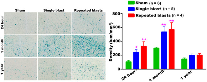
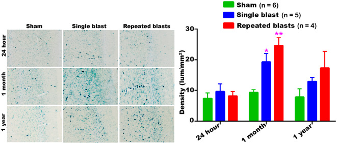
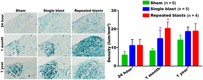
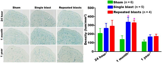
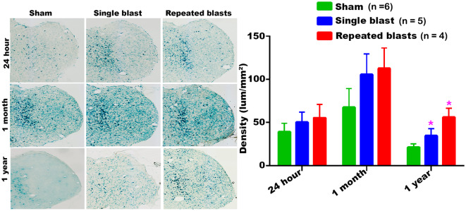
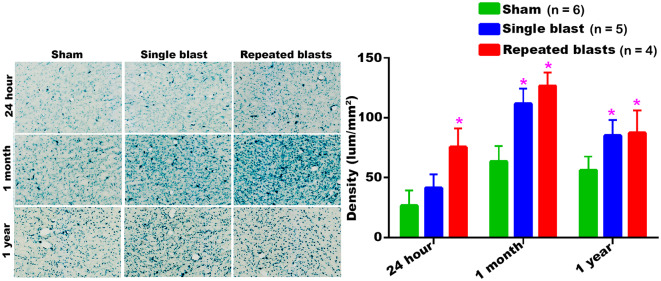
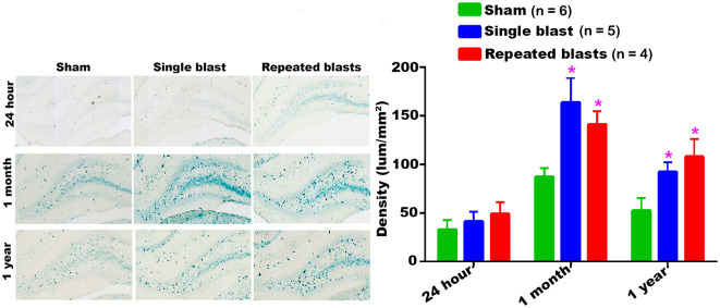
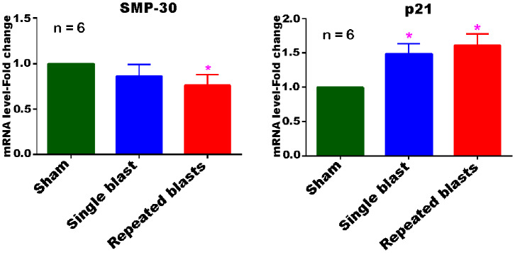
Blast-induced traumatic brain injury (bTBI) is one of the major causes of persistent disabilities in Service Members, and a history of bTBI has been identified as a primary risk factor for developing age-associated neurodegenerative diseases. Clinical observations of several military blast casualties have revealed a rapid age-related loss of white matter integrity in the brain. In the present study, we have tested the effect of single and tightly coupled repeated blasts on cellular senescence in the rat brain. Isoflurane-anesthetized rats were exposed to either a single or 2 closely coupled blasts in an advanced blast simulator. Rats were euthanized and brains were collected at 24 h, 1 month and 1 year post-blast to determine senescence-associated-β-galactosidase (SA-β-gal) activity in the cells using senescence marker stain. Single and repeated blast exposures resulted in significantly increased senescence marker staining in several neuroanatomical structures, including cortex, auditory cortex, dorsal lateral thalamic nucleus, geniculate nucleus, superior colliculus, ventral thalamic nucleus and hippocampus. In general, the increases in SA-β-gal activity were more pronounced at 1 month than at 24 h or 1 year post-blast and were also greater after repeated than single blast exposures. Real-time quantitative RT-PCR analysis revealed decreased levels of mRNA for senescence marker protein-30 (SMP-30) and increased mRNA levels for p21 (cyclin dependent kinase inhibitor 1A, CDKN1A), two other related protein markers of cellular senescence. The increased senescence observed in some of these affected brain structures may be implicated in several long-term sequelae after exposure to blast, including memory disruptions and impairments in movement, auditory and ocular functions.

Keywords: SA-β-gal; SMP-30; aging; blast exposure; p21; senescence; traumatic brain injury.


Figures

Figure 1
Figure 1
Activity of SA-β-gal in the motor cortex at different intervals post-blast exposures. Density values are expressed as mean ± SEM. Values of all three groups were compared to each other at each time point for statistical significant differences. *Blast exposed groups at each time point were compared to corresponding sham controls (*p < 0.05; **p < 0.01; n = 4–6).
Figure 2
Figure 2
SA-β-gal activity in the auditory cortex at different intervals post-blast exposures. Density values are expressed as mean ± SEM. Values of all three groups were compared to each other at each time point for statistical significance. *Blast exposed groups at each time point were compared to corresponding sham controls (*p < 0.05; **p < 0.01; n = 4–6).
Figure 3
Figure 3
Dorsolateral thalamus showing the differential activity of SA-β-gal at different intervals post-blast exposures. Density values are expressed as mean ± SEM. Values of all three groups were compared to each other at each time point for statistical significance. *Blast exposed groups at each time point were compared to corresponding sham controls (*p < 0.05; n = 4–6).
Figure 4
Figure 4
Activity of SA-β-gal in the superior colliculus at different intervals post-blast exposures. Density values are expressed as mean ± SEM. Values of all three groups were compared to each other at each time point for statistical significance. *Blast exposed groups at each time point were compared to corresponding sham controls (*p < 0.05; n = 4–6).
Figure 5
Figure 5
Geniculate nucleus showing the differential activity of SA-β-gal at different intervals post-blast exposures. Density values are expressed as mean ± SEM. Values of all three groups were compared to each other at each time point for statistical significance. *Blast exposed groups at each time point were compared to corresponding sham controls (*p < 0.05; n = 4–6).
Figure 6
Figure 6
SA-β-gal activity in the ventral thalamic nucleus at different intervals post-blast exposures. Density values are expressed as mean ± SEM. Values of all three groups were compared to each other at each time point for statistical significance. *Blast exposed groups at each time point were compared to corresponding sham controls (*p < 0.05; n = 4–6).
Figure 7
Figure 7
Activity of SA-β-gal in the hippocampus at different intervals post-blast exposures. Density values are expressed as mean ± SEM. Values of all three groups were compared to each other at each time point for statistical significance. *Blast exposed groups at each time point were compared to corresponding sham controls (*p < 0.05; n = 4–6).
Figure 8
Figure 8
Differential expression of SMP-30 and p21 mRNAs in the cerebral cortex at 1 month post-blast exposures. Values of the blast exposed groups are expressed as mean ± SEM. Fold changes in the expressions of mRNAs of blast exposed groups were compared to those of sham controls (*p < 0.05; n = 6).

References

    1. Reid MW, Miller KJ, Lange RT, Cooper DB, Tate DF, Bailie J, et al. A multisite study of the relationships between blast exposures and symptom reporting in a post-deployment active duty military population with mild traumatic brain injury. J Neurotrauma. (2014) 31:1899–906. 10.1089/neu.2014.3455 - DOI - PMC - PubMed
    1. Warden D. Military TBI during the Iraq and Afghanistan wars. J Head Trauma Rehabil. (2006) 21:398–402. 10.1097/00001199-200609000-00004 - DOI - PubMed
    1. Kontos AP, Elbin RJ, Kotwal RS, Lutz RH, Kane S, Benson PJ, et al. The effects of combat-related mild traumatic brain injury (mTBI): does blast mTBI history matter? J Trauma Acute Care Surg. (2015) 79:S146–51. 10.1097/TA.0000000000000667 - DOI - PubMed
    1. Fear NT, Jones E, Groom M, Greenberg N, Hull L, Hodgetts TJ, et al. Symptoms of post-concussional syndrome are non-specifically related to mild traumatic brain injury in UK armed forces personnel on return from deployment in Iraq: an analysis of self-reported data. Psychol Med. (2009) 39:1379–87. 10.1017/S0033291708004595 - DOI - PubMed
    1. Levin HS, Wilde E, Troyanskaya M, Petersen NJ, Scheibel R, Newsome M, et al. Diffusion tensor imaging of mild to moderate blast-related traumatic brain injury and its sequelae. J Neurotrauma. (2010) 27:683–94. 10.1089/neu.2009.1073 - DOI - PubMed