. 2020 May 19:11:864.
doi: 10.3389/fimmu.2020.00864. eCollection 2020.

Contrasting Inflammatory Signatures in Peripheral Blood and Bronchoalveolar Cells Reveal Compartment-Specific Effects of HIV Infection

Affiliations

Contrasting Inflammatory Signatures in Peripheral Blood and Bronchoalveolar Cells Reveal Compartment-Specific Effects of HIV Infection

Daniel M Muema et al. Front Immunol. .

Abstract

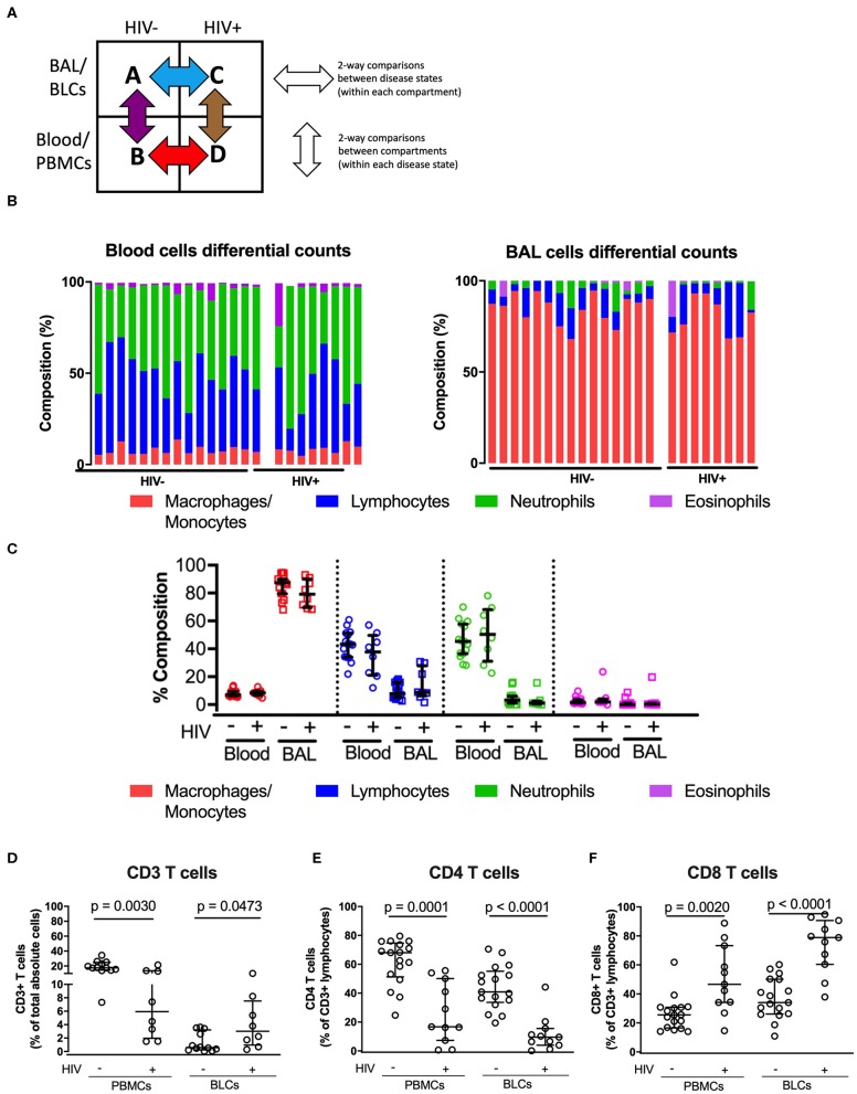
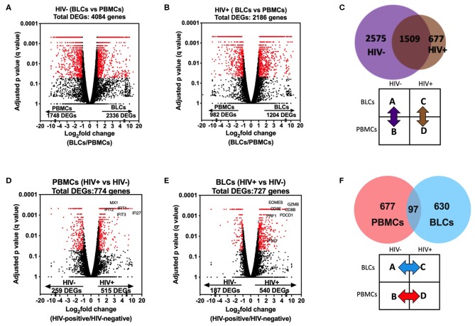
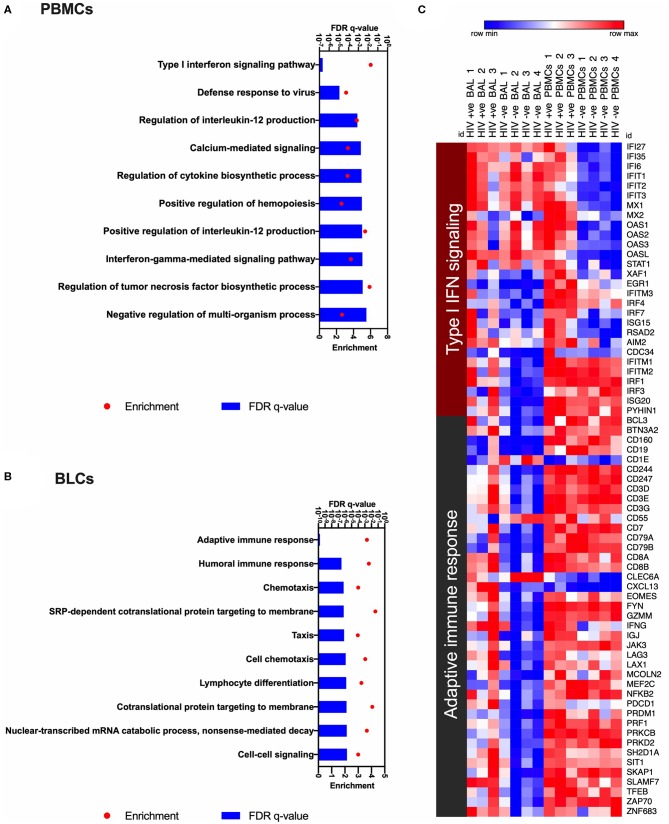
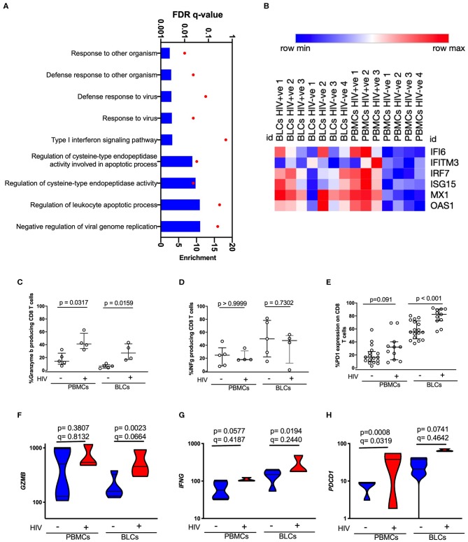
The mechanisms by which HIV increases susceptibility to tuberculosis and other respiratory infections are incompletely understood. We used transcriptomics of paired whole bronchoalveolar lavage cells (BLCs) and peripheral blood mononuclear cells to compare the effect of HIV at the lung mucosal surface and in peripheral blood. The majority of HIV-induced differentially expressed genes (DEGs) were specific to either the peripheral or lung mucosa compartments (1,307/1,404, 93%). Type I interferon signaling was the dominant signature of DEGs in HIV-positive blood but not in HIV-positive BLCs. DEGs in the HIV-positive BLCs were significantly enriched for infiltration with cytotoxic CD8+ T cells. Higher expression of type 1 interferon transcripts in peripheral CD8+ T cells and representative transcripts and proteins in BLCs-derived CD8+ T cells during HIV infection, including IFNG (IFN-gamma), GZMB (Granzyme B), and PDCD1 (PD-1), was confirmed by cell-subset specific transcriptional analysis and flow cytometry. Thus, we report that a whole transcriptomic approach revealed qualitatively distinct effects of HIV in blood and bronchoalveolar compartments. Further work exploring the impact of distinct type I interferon programs and functional features of CD8+ T cells infiltrating the lung mucosa during HIV infection may provide novel insights into HIV-induced susceptibility to respiratory pathogens.

Keywords: Cytotoxic CD8 T cells; HIV; bronchoalveolar; lymphocyte infiltration; type I interferon.


Figures

Figure 1
Figure 1
Comparisons of the immune cell sub-types between peripheral blood and bronchoalveolar lavage (BAL) cells of both HIV-negative and HIV-positive individuals. (A) Two-way comparison of the study groups to understand inter-compartment differences and the impact of HIV on immune function. (B) Proportion of immune cell subsets in the paired peripheral blood and bronchoalveolar cells in the HIV-negative (n = 15) and HIV-positive groups (n = 8). (C) Quantitative comparison of panel (B), showing level of significance between compartments in both HIV-negative and HIV-positive groups. (D) Proportions of total absolute CD3+ T cells in BLCs and PBMCs in HIV-positive (n = 8) and HIV-negative groups (n = 15). (E) Percentage CD4+ T cells of CD3+ T cells in BLCs and PBMCs in HIV-positive (n = 11) and HIV-negative groups (n = 19). (F) Percentage of CD8+ T cells in BLCs and PBMCs in HIV-positive (n = 11) and HIV-negative groups (n = 19).
Figure 2
Figure 2
Comparisons in transcriptomic profiles between bronchoalveolar lavage fluid cells (BLCs) and peripheral blood mononuclear cells (PBMCs) in HIV-infected and HIV-uninfected participants. (A) Differentially expressed genes between BLCs and PBMCs in the HIV-negative group (n = 4). (B) Differentially expressed genes between BLCs and PBMCs in the HIV-positive group (n = 3). (C) Numbers of differentially expressed genes between BLCs and PBMCs in both HIV-positive (n = 3) and HIV-negative groups (n = 4). (D) Differentially expressed genes between HIV-negative (n = 4) and HIV-positive (n = 3) groups in PBMCs. (E) Differentially expressed genes between HIV-negative and HIV-positive groups in BLCs. (F) Numbers of differentially expressed genes between HIV-negative and HIV-positive groups in both PBMCs and BLCs.
Figure 3
Figure 3
Gene ontology analyses to identify HIV-induced enrichments in bronchoalveolar lavage fluid cells (BLCs) and peripheral blood mononuclear cells (PBMCs). (A) The top 10 HIV-associated significantly enriched gene ontology (GO) terms in PBMCs. (B) The top 10 HIV-associated significantly enriched gene ontology (GO) terms in BLCs. (C) Heat map showing expression levels of the genes that contribute to the most enriched GO terms in PBMCs and BLCs, i.e., “Type I interferon signaling pathway” and “Adaptive immune response.” Natural logarithms of (Expression level +1) were used.
Figure 4
Figure 4
Characterization of HIV-induced changes in PBMCs-derived CD8 T cells and BLCs-derived CD8 T cells. (A) Enriched gene ontology (GO) terms in PBMCs-derived CD8+ T cells in comparisons between HIV-positive individuals and HIV-negative individuals. (B) Heat map showing expression levels of type I interferon inducible genes in sorted CD8+ T cells. Natural logarithms of (Expression level +1) were used. (C) Constitutive expression of granzyme B in unstimulated PBMCs-derived CD8 T cells and BLCs-derived CD8+ T cells from HIV-positive and HIV-negative participants. (D) Inducible interferon gamma in PBMCs-derived CD8+ T cells and BLCs-derived CD8 T cells from HIV-positive and HIV-negative participants. (E) Ex vivo expression of PD-1 in PBMCs-derived CD8+ T cells and BLCs-derived CD8 T cells in HIV-positive and HIV-negative participants. (F) Expression levels of granzyme B mRNA (GZMB) in BLCs-derived sorted CD8+ T cells and PBMCs-derived sorted CD8+ T cells from HIV-positive and HIV-negative participants. (G) Constitutive expression levels of interferon gamma mRNA (IFNG) in BLCs-derived sorted CD8+ T cells and PBMCs-derived sorted CD8+ T cells from HIV-positive and HIV-negative participants. (H) Expression levels of PD-1 mRNA (PDCD1) in BLCs-derived and PBMCs-derived sorted CD8+ T cells from HIV-positive and HIV-negative participants.

References

    1. UNAIDS DATA. (2019). Available online at: https://www.unaids.org/sites/default/files/media_asset/2019-UNAIDS-data_... (accessed September 4, 2019).
    1. UNAIDS Tuberculosis and HIV. (2019). Available online at: https://www.unaids.org/sites/default/files/media_asset/tuberculosis-and-... (accessed May 6, 2019).
    1. Fenner L, Atkinson A, Boulle A, Fox MP, Prozesky H, Zurcher K, et al. . HIV viral load as an independent risk factor for tuberculosis in South Africa: collaborative analysis of cohort studies. J Int AIDS Soc. (2017) 20:21327. 10.7448/IAS.20.1.21327 - DOI - PMC - PubMed
    1. Caruso AM, Serbina N, Klein E, Triebold K, Bloom BR, Flynn JL. Mice deficient in CD4 T cells have only transiently diminished levels of IFN-γ, yet succumb to tuberculosis. J Immunol. (1999) 162:5407–16. - PubMed
    1. Flynn JL, Chan J, Triebold KJ, Dalton DK, Stewart TA, Bloom BR. An essential role for interferon gamma in resistance to Mycobacterium tuberculosis infection. J Exp Med. (1993) 178:2249–54. 10.1084/jem.178.6.2249 - DOI - PMC - PubMed

Publication types

MeSH terms