Review
. 2020 May 21:11:924.
doi: 10.3389/fimmu.2020.00924. eCollection 2020.

Dendritic Cells and Their Role in Immunotherapy

Affiliations
Review

Dendritic Cells and Their Role in Immunotherapy

Alycia Gardner et al. Front Immunol. .

Abstract

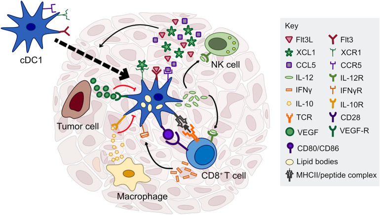
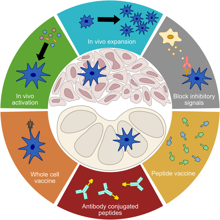
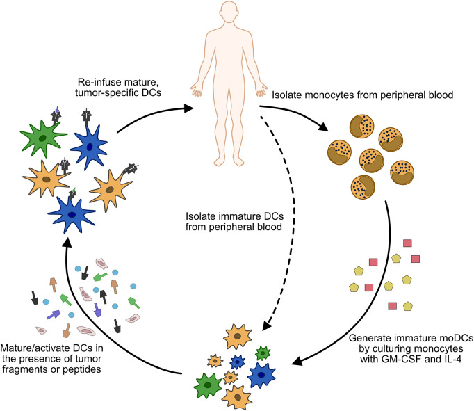
Despite significant advances in the field of cancer immunotherapy, the majority of patients still do not benefit from treatment and must rely on traditional therapies. Dendritic cells have long been a focus of cancer immunotherapy due to their role in inducing protective adaptive immunity, but cancer vaccines have shown limited efficacy in the past. With the advent of immune checkpoint blockade and the ability to identify patient-specific neoantigens, new vaccines, and combinatorial therapies are being evaluated in the clinic. Dendritic cells are also emerging as critical regulators of the immune response within tumors. Understanding how to augment the function of these intratumoral dendritic cells could offer new approaches to enhance immunotherapy, in addition to improving the cytotoxic and targeted therapies that are partially dependent upon a robust immune response for their efficacy. Here we will discuss the role of specific dendritic cell subsets in regulating the anti-tumor immune response, as well as the current status of dendritic cell-based immunotherapies, in order to provide an overview for future lines of research and clinical trials.

Keywords: cancer; dendritic cells; immune checkpoint blockade; immunotherapy; vaccines.


Figures

Figure 1
Figure 1
Factors regulating cDC1 function in the tumor microenvironment. cDC1s interact with several immune cell types through cytokine and chemokine signaling, including NK cells, T cells, and macrophages. NK cells are critical for cDC1 recruitment and survival in the tumor through production of Flt3L, CCL5, and XCL1. cDC1 have the capacity to cross-present exogenous antigen to CD8+ T cells and stimulate naïve and previously activated T cells ex vivo; however, the importance of antigen presentation by cDC1s in the tumor microenvironment is currently unclear. In contrast, cDC1 production of IL-12, driven by IFN-γ or other inflammatory mediators, is necessary to sustain a T cell response during chemotherapy or immune checkpoint blockade. cDC1 production of IL-12 can be directly inhibited by IL-10 released by macrophages or other immunosuppressive cells, as well as tumor-derived factors that inhibit the maturation of cDC1s such as VEGF.
Figure 2
Figure 2
Treatment modalities targeting DCs. A number of current treatment modalities aim to address limited DC functionality in order to elicit or enhance anti-tumor immune responses. Treatments that seek to improve the function of tumor DCs include in vivo activation, in vivo expansion, and the blocking of inhibitory signals. Vaccination approaches that seek to bypass tumor DCs and directly stimulate a de novo T cell response in the lymph nodes include whole cell vaccines, antibody conjugated peptides, and free proteins or peptides.
Figure 3
Figure 3
Process of generating whole cell DC vaccines. Monocytes (or less commonly, immature cDCs) are isolated from the patient's peripheral blood. In the case of monocyte isolation, immature moDCs are generated by culturing the isolated cells in GM-CSF and IL-4. Once immature DCs are obtained, they are matured/activated using a variety of cytokine cocktails, and pulsed with tumor antigen or tumor fragments. The matured DCs are then injected back into the patient, usually via subcutaneous (s.c.) or intradermal (i.d.) injections, although intravenous (i.v.) or direct injection into lymph nodes has also been used.

References

    1. Chen DS, Mellman I. Oncology meets immunology: the cancer-immunity cycle. Immunity. (2013) 39:1–10. 10.1016/j.immuni.2013.07.012 - DOI - PubMed
    1. Nakayama M. Antigen presentation by MHC-Dressed cells. Front Immunol. (2014) 5:672. 10.3389/fimmu.2014.00672 - DOI - PMC - PubMed
    1. Lin A, Lore K. Granulocytes: new members of the antigen-presenting cell family. Front Immunol. (2017) 8:1781. 10.3389/fimmu.2017.01781 - DOI - PMC - PubMed
    1. Broz ML, Binnewies M, Boldajipour B, Nelson AE, Pollack JL, Erle DJ, et al. Dissecting the tumor myeloid compartment reveals rare activating antigen-presenting cells critical for T cell immunity. Cancer Cell. (2014) 26:638–52. 10.1016/j.ccell.2014.09.007 - DOI - PMC - PubMed
    1. DeNardo DG, Ruffell B. Macrophages as regulators of tumour immunity and immunotherapy. Nat Rev Immunol. (2019) 19:369–82. 10.1038/s41577-019-0127-6 - DOI - PMC - PubMed

Publication types

MeSH terms

Substances