Review
. 2020 May 26;6(1):32.
doi: 10.1038/s41421-020-0161-3. eCollection 2020.

Emerging roles of ATG proteins and membrane lipids in autophagosome formation

Affiliations
Review

Emerging roles of ATG proteins and membrane lipids in autophagosome formation

Taki Nishimura et al. Cell Discov. .

Abstract

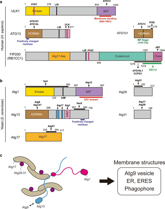
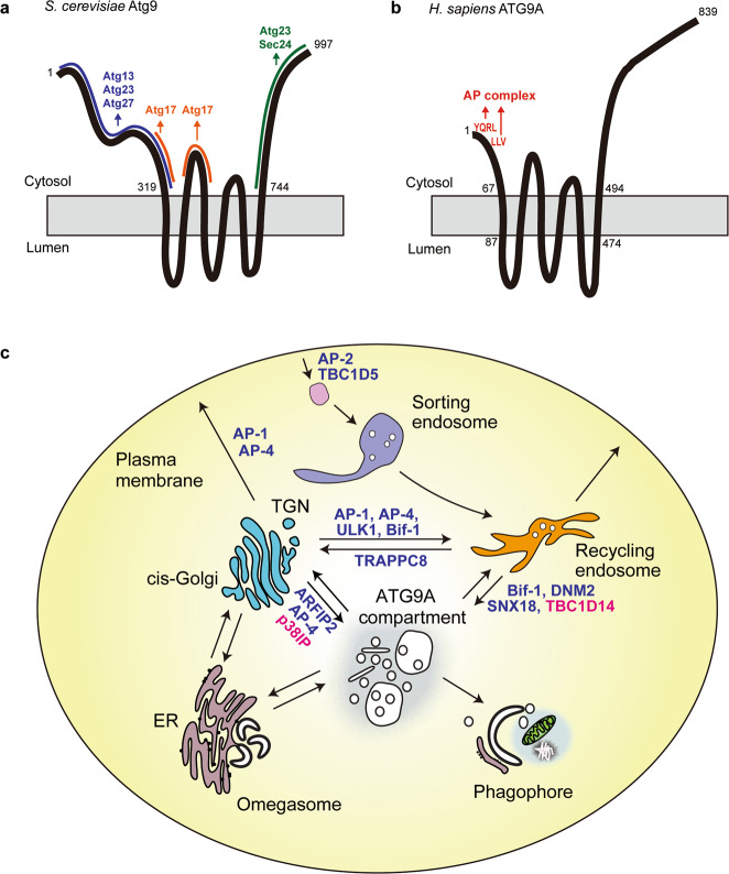
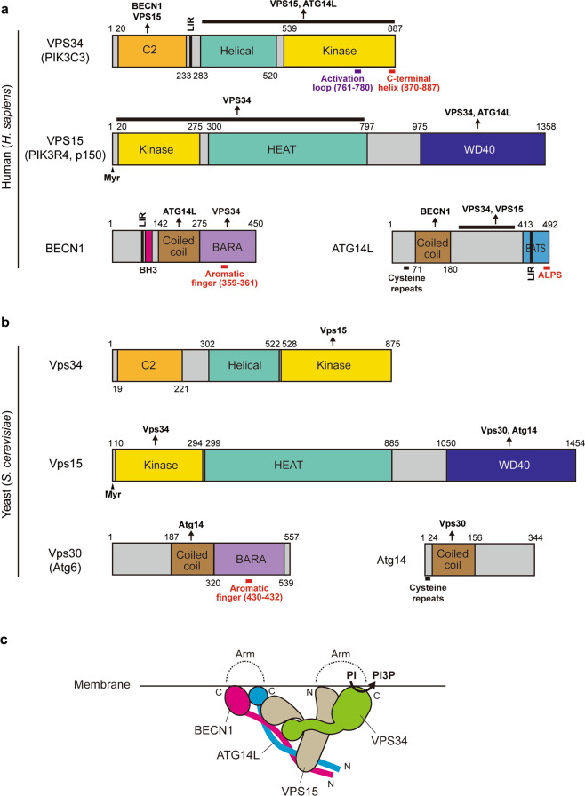
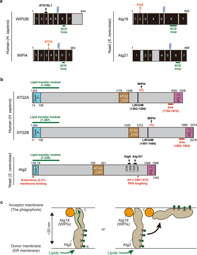
Autophagosome biogenesis is a dynamic membrane event, which is executed by the sequential function of autophagy-related (ATG) proteins. Upon autophagy induction, a cup-shaped membrane structure appears in the cytoplasm, then elongates sequestering cytoplasmic materials, and finally forms a closed double membrane autophagosome. However, how this complex vesicle formation event is strictly controlled and achieved is still enigmatic. Recently, there is accumulating evidence showing that some ATG proteins have the ability to directly interact with membranes, transfer lipids between membranes and regulate lipid metabolism. A novel role for various membrane lipids in autophagosome formation is also emerging. Here, we highlight past and recent key findings on the function of ATG proteins related to autophagosome biogenesis and consider how ATG proteins control this dynamic membrane formation event to organize the autophagosome by collaborating with membrane lipids.

Keywords: Endoplasmic reticulum; Macroautophagy.


Conflict of interest statement

Conflict of interestThe authors declare that they have no conflict of interest.

Figures

Fig. 1
Fig. 1. ATG/Atg proteins control dynamic membrane events during autophagosome biogenesis.
a Autophagosome formation can be dissected into five different steps: initiation, nucleation, membrane expansion, closure, and fusion. b, c The intracellular distribution of ATG/Atg proteins under starvation-induced autophagy in mammalian (b),–,,,,,,, and yeast cells (c),,,,,,,. Their localizations are categorized into five groups: -, not detectable; -/+, transient; +, weakly detectable; ++, easily detectable; +++, clearly detectable. Note that ATG/Atg proteins show punctate structures on the ER-related membranes rather than a typical ER-like pattern. ERES ER exit sites, ERGIC ER-Golgi intermediate compartment.
Fig. 2
Fig. 2. The ULK/Atg1 complex is recruited to membrane structures to initiate autophagy.
a The domain structures of H. sapiens ULK complex components. b The domain structures of S. cerevisiae Atg1 complex components. c The proposed structure of the Atg1 complex. EAT early autophagy targeting and tethering, MIT microtubule interacting and transport, MIM MIT-interacting motif, LIR LC3-interacting region, AIM Atg8 family-interacting motif, FFAT two phenylalanines in an acidic tract, HORMA Hop1/Rev7/Mad2.
Fig. 3
Fig. 3. Atg9/ATG9A vesicles work as a membrane source for autophagosome formation.
a, b The domain structure of S. cerevisiae Atg9 (a) and H. sapiens ATG9A (b) proteins. c Mammalian ATG9A cycles between different organelle compartments via vesicular transport pathways. Positive and negative regulators are shown in blue and red, respectively. AP adaptor protein, ARFIP2 arfaptin-2, SNX18 sorting nexin 18, DNM2 dynamin 2, TBC1D14 TBC1 domain family member 14, TRAPPC8 trafficking protein particle complex 8, p38IP p38-interacting protein.
Fig. 4
Fig. 4. The class III PI3K complex I (PI3KC3-C1) synthesizes PI3P at the autophagy initiation site.
a, b The domain structures of H. sapiens PI3KC3-C1 components (a) and S. cerevisiae PI3KC3-C1 components (b). c The proposed structure of mammalian PI3KC3-C1 complex. HEAT, Huntingtin, elongation factor 3, the PR65/A subunit of protein phosphatase 2A and the lipid kinase Tor; BH3 Bcl-2 homology 3, LIR LC3-interacting region, BARA β-α repeated autophagy-specific, Myr Myristoylation, BATS BAKOR and ATG14L autophagosome-targeting sequence, ALPS amphipathic lipid packing sensor.
Fig. 5
Fig. 5. Atg2/ATG2 functions as a membrane tether and lipid transfer protein in autophagy.
a The domain structures of WIPI2B and WIPI4 in human (left) and Atg18 and Atg21 in S. cerevisiae (right). b The domain structures of Atg2/ATG2 proteins. c Models of Atg2/ATG2-dependent lipid transfer. Chorein_N N-terminal region of Chorein or VPS13, Atg2_CAD autophagy-related protein 2 CAD motif, ATG_C autophagy-related protein C-terminal domain, LIR/GIM LC3/GABARAP-interacting region, AH amphipathic helix.
Fig. 6
Fig. 6. Atg12–5-16/ATG12–5-16L1 and Atg3/ATG3 catalyze lipidation of Atg8/LC3 family proteins.
a The lipidation system of LC3. ATG4 cleaves the C-terminal residues of LC3 to expose glycine (G) residue. Then, LC3 is activated by ATG7 (E1 enzyme) and transferred to ATG3 (E2 enzyme). ATG12–5-16L1 complex facilitates the transfer of LC3 from ATG3 to PE. A PI3P-binding protein WIPI2B controls membrane recruitment of ATG12–5-16L1 complex under starvation condition. b The domain structures of H. sapiens ATG16L1 and S. cerevisiae Atg16 proteins. c The domain structures of H. sapiens ATG3 and S. cerevisiae Atg3 proteins. CC coiled-coil, AH amphipathic helix, FR flexible region, HR handle region, AIM Atg8 family-interacting motif.

References

    1. Dunn WA., Jr. Studies on the mechanisms of autophagy: formation of the autophagic vacuole. J. Cell Biol. 1990;110:1923–1933. doi: 10.1083/jcb.110.6.1923. - DOI - PMC - PubMed
    1. Deter RL, Baudhuin P, De Duve C. Participation of lysosomes in cellular autophagy induced in rat liver by glucagon. J. Cell Biol. 1967;35:C11–C16. doi: 10.1083/jcb.35.2.C11. - DOI - PMC - PubMed
    1. Novikoff AB, Shin WY. Endoplasmic reticulum and autophagy in rat hepatocytes. Proc. Natl Acad. Sci. USA. 1978;75:5039–5042. doi: 10.1073/pnas.75.10.5039. - DOI - PMC - PubMed
    1. Kopitz J, Kisen GO, Gordon PB, Bohley P, Seglen PO. Nonselective autophagy of cytosolic enzymes by isolated rat hepatocytes. J. Cell Biol. 1990;111:941–953. doi: 10.1083/jcb.111.3.941. - DOI - PMC - PubMed
    1. Stromhaug PE, Berg TO, Fengsrud M, Seglen PO. Purification and characterization of autophagosomes from rat hepatocytes. Biochem. J. 1998;335(Pt 2):217–224. doi: 10.1042/bj3350217. - DOI - PMC - PubMed