Case Reports
. 2020 May 14:2020:6723936.
doi: 10.1155/2020/6723936. eCollection 2020.

Horizontal Ridge Augmentation Using a Xenograft Bone Substitute for Implant-Supported Fixed Rehabilitation: A Case Report with Four Years of Follow-Up

Affiliations
Case Reports

Horizontal Ridge Augmentation Using a Xenograft Bone Substitute for Implant-Supported Fixed Rehabilitation: A Case Report with Four Years of Follow-Up

Bruno Freitas Mello et al. Case Rep Dent. .

Abstract

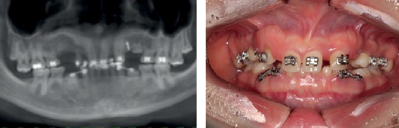
The guided bone regeneration (GBR) technique has been used to achieve optimal bone volume augmentation and allow dental implant placement in atrophic maxilla and mandible, with predictable results and high survival rates. The use of bone substitutes has reduced the necessity of autogenous bone grafts, reducing the morbidity at the donor areas and thus improving the patients' satisfaction and comfort. This clinical case report shows a clinical and histological evaluation of the bone tissue behavior, in a case that required the horizontal augmentation of the alveolar ridge, with the use of xenograft biomaterial and further dental implant placement. After six months of healing time, six implants were placed, and a bone biopsy was done. The histological analysis depicted some fragments of the xenograft bone graft, integrated with the new-formed bone tissue.


Conflict of interest statement

The authors declare that there is no conflict of interest regarding the publication of this paper.

Figures

Figure 1
Figure 1
Panoramic radiograph (a) and intraoral buccal view, showing permanent tooth absences and presence of several deciduous teeth.
Figure 2
Figure 2
Preoperatory tomography of the anterior mandible region.
Figure 3
Figure 3
Intraoral view of the surgery steps: (a) incision and total flap detachment, (b) cortical perforations, (c) bone substitute accommodation, (d) double layer collagen membrane, (e) single sutures, and (f) bandage (Micropore®).
Figure 4
Figure 4
Postoperatory tomography radiograph 12 months after the horizontal bone augmentation, showing new bone formation, allowing the correct three-dimensional placement of dental implants.
Figure 5
Figure 5
3D resin prototype of the appearance of the ridge before (a) and after (b) the volume increase of the alveolar ridge. There was a 5 mm gain in bone thickness.
Figure 6
Figure 6
Intraoral view of the alveolar ridge before (a) and after (b) augmentation. Removal of bone fragment using a 2.0 mm trephine for histological analysis of the grafted area (c). Six implants in place with initial torque of 35 N/cm (d).
Figure 7
Figure 7
(a) Image of a grafted material interface (red arrow) and cortical bone. It is clearly shown the formation of cortical bone with osteocytes; (b) enlarged image of (a). Grafted material (red arrow). Osteocytes present in the gaps (green arrow); (c) well-formed Haversian system: note the arrangement of concentric lamellae, with the central channel of Havers (blue arrow). Osteocytes homogeneously distributed, demonstrating the usual architecture of cortical bone; (c) enlarged image of (d). In detail, the Havers channel, with endosteum (orange arrow) and red blood cells (white arrow).
Figure 8
Figure 8
Installation of the definitive prosthetic abutments and delivery of the temporary cemented restoration with immediate load.
Figure 9
Figure 9
Four-year follow-up CT, showing the stability of the bone tissue around the implants (a, b). Panoramic radiograph of the 4-year follow-up (c).
Figure 10
Figure 10
Buccal view of the four-year follow-up.

References

    1. Al-Nawas B., Schiegnitz E. Augmentation procedures using bone substitute materials or autogenous bone – a systematic review and meta-analysis. European Journal of Oral Implantology. 2014;7(Supplement 2):S219–S234. - PubMed
    1. Chiapasco M., Zaniboni M. Clinical outcomes of GBR procedures to correct peri-implant dehiscences and fenestrations: a systematic review. Clinical Oral Implants Research. 2009;20(Supplement 4):113–123. doi: 10.1111/j.1600-0501.2009.01781.x. - DOI - PubMed
    1. Urban I. A., Jovanovic S. A., Lozada J. L. Vertical ridge augmentation using guided bone regeneration (GBR) in three clinical scenarios prior to implant placement: a retrospective study of 35 patients 12 to 72 months after loading. The International Journal of Oral & Maxillofacial Implants. 2009;24(3):502–510. - PubMed
    1. Aghaloo T. L., Moy P. K. Which hard tissue augmentation techniques are the most successful in furnishing bony support for implant placement? The International Journal of Oral & Maxillofacial Implants. 2007;22:49–70. - PubMed
    1. Retzepi M., Donos N. Guided bone regeneration: biological principle and therapeutic applications. Clinical Oral Implants Research. 2010;21(6):567–576. doi: 10.1111/j.1600-0501.2010.01922.x. - DOI - PubMed

Publication types

LinkOut - more resources