

Response Evaluation of Choroidal Melanoma After Brachytherapy Using Diffusion-Weighted Magnetic Resonance Imaging (DW-MRI): Preliminary Findings
- PMID: 32509587
- PMCID: PMC7248391
- DOI: 10.3389/fonc.2020.00825
Response Evaluation of Choroidal Melanoma After Brachytherapy Using Diffusion-Weighted Magnetic Resonance Imaging (DW-MRI): Preliminary Findings
Abstract
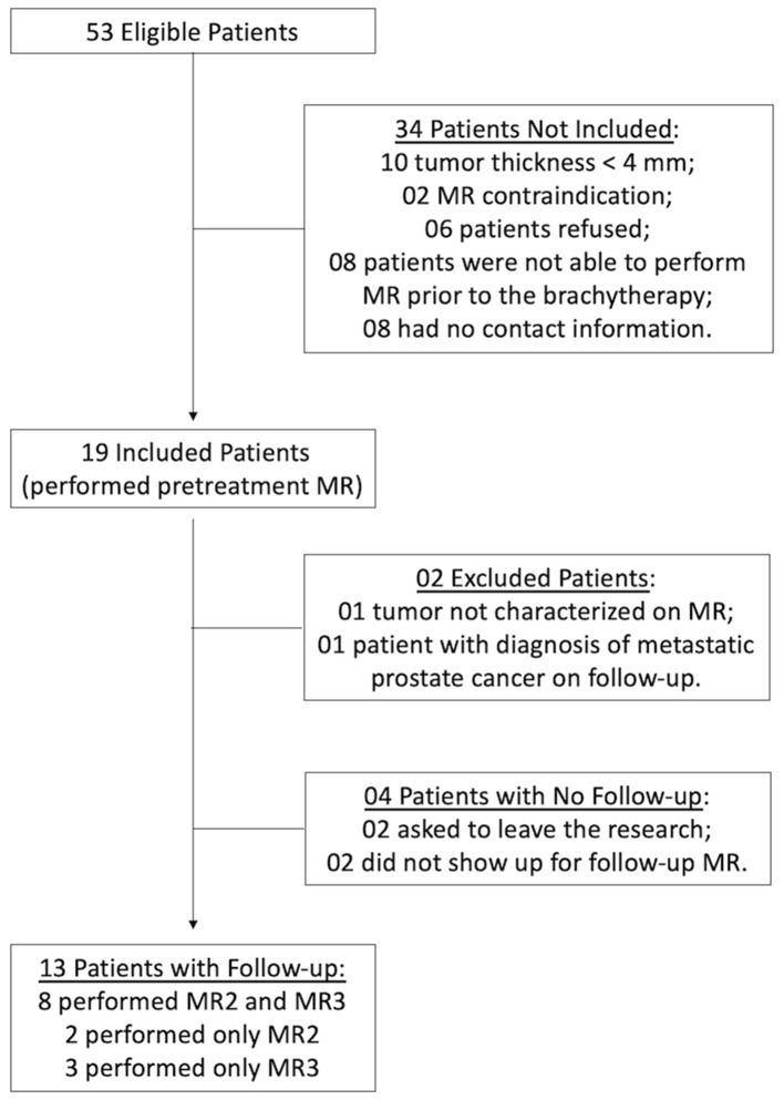
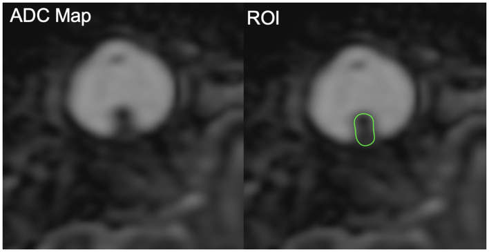
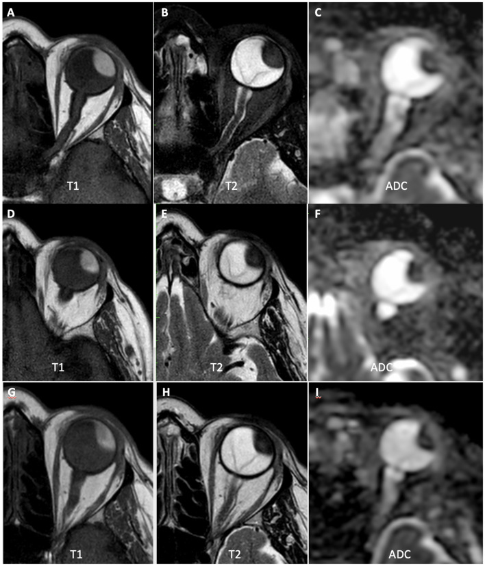
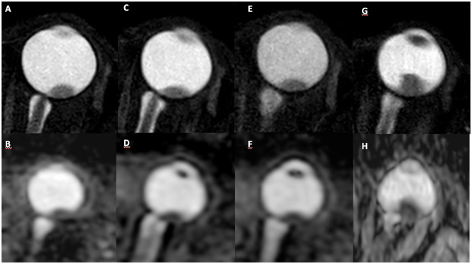
Purpose: To evaluate the role of diffusion-weighted magnetic resonance imaging (DW-MRI) in the assessment of therapeutic response in patients with choroidal melanoma treated with brachytherapy. Materials and Methods: We performed a prospective, unicentric study which included patients with choroidal melanoma and indication for brachytherapy. Three DW-MRI examinations were proposed for each patient, one before and two after treatment. The apparent diffusion coefficient (ADC) value was calculated on DW-MRI and compared with local tumor control assessed by ophthalmologic follow-up. Results: From 07/2018 to 06/2019, 19 patients were recruited, 13 of whom underwent follow-up examinations. Patients' ages ranged from 24 to 78 years and 52.9% were male. At the ocular ultrasound, the mean tumor thickness and diameter were 6.3 and 11.5 mm, respectively. Two patients (15.4%) showed signs of tumor progression during follow-up (7 and 9 months after treatment). There was no statistically significant difference in tumor size between MR before and after treatment, however, there was a significant reduction in mean ADC in patients with progression (p = 0.02). Conclusion: DW-MRI is a promising method for monitoring patients with choroidal melanoma; reduction in the mean ADC values between pre-treatment MRI and the first post-treatment MRI may be related to the lack of response to brachytherapy and increased risk of disease progression.
Keywords: brachytherapy; diffusion magnetic resonance imaging; eye neoplasms; melanoma; response evaluation criteria in solid tumors.
Copyright © 2020 Bitencourt, Bitencourt, Chojniak, Souza, Castro, Pellizzon and Chojniak.
Figures
Similar articles
-
PET/CT in brachytherapy early response evaluation of pancreatic ductal adenocarcinoma xenografts: comparison with apparent diffusion coefficient from diffusion-weighted MR imaging.Abdom Radiol (NY). 2019 Mar;44(3):950-957. doi: 10.1007/s00261-018-1791-x. Abdom Radiol (NY). 2019. PMID: 30315322
-
Coronal diffusion-weighted magnetic resonance imaging of the kidney: agreement with axial diffusion-weighted magnetic imaging in terms of apparent diffusion coefficient values.Chin Med J (Engl). 2015 Feb 20;128(4):499-503. doi: 10.4103/0366-6999.151103. Chin Med J (Engl). 2015. PMID: 25673453 Free PMC article. Clinical Trial.
-
Diffusion-weighted MRI Before and After Robotic Radiosurgery (Cyberknife®) in Primary and Secondary Liver Malignancies: A Pilot Study.Technol Cancer Res Treat. 2015 Apr;14(2):191-9. doi: 10.7785/tcrt.2012.500408. Epub 2014 Nov 21. Technol Cancer Res Treat. 2015. PMID: 24502549
-
Diffusion-weighted magnetic resonance imaging and ultrasound evaluation of choroidal melanomas after proton-beam therapy.Radiol Med. 2015 Jul;120(7):634-40. doi: 10.1007/s11547-015-0509-1. Epub 2015 Feb 4. Radiol Med. 2015. PMID: 25650084
-
Magnetic resonance diffusion-weighted imaging: extraneurological applications.Radiol Med. 2006 Apr;111(3):392-419. doi: 10.1007/s11547-006-0037-0. Epub 2006 Apr 11. Radiol Med. 2006. PMID: 16683086 Review. English, Italian.
Cited by
-
Radiological and clinical findings in uveal melanoma treated by plaque interventional radiotherapy (brachytherapy): Visual atlas and literature review on response assessment.J Contemp Brachytherapy. 2022 Feb;14(1):96-106. doi: 10.5114/jcb.2022.113271. Epub 2022 Feb 4. J Contemp Brachytherapy. 2022. PMID: 35233241 Free PMC article.
-
Magnetic Resonance Imaging in the Clinical Care for Uveal Melanoma Patients-A Systematic Review from an Ophthalmic Perspective.Cancers (Basel). 2023 May 30;15(11):2995. doi: 10.3390/cancers15112995. Cancers (Basel). 2023. PMID: 37296958 Free PMC article. Review.
References
-
- Chojniak MM, Erwenne CM. Braquiterapia com Cobalto 60 para o tratamento do melanoma da úvea: Análise dos fatores prognósticos para melhor resposta local. Arq Bras Oftalmol. (2002) 65:199–206. 10.1590/S0004-27492002000200008 - DOI
LinkOut - more resources
Full Text Sources