

Spontaneous Necrosis of a Large Choroidal Melanoma: Unusual Presentation in a 49-Year-Old Male
- PMID: 32509762
- PMCID: PMC7250329
- DOI: 10.1159/000501522
Spontaneous Necrosis of a Large Choroidal Melanoma: Unusual Presentation in a 49-Year-Old Male
Abstract
Purpose: To demonstrate a case of massive vitreous haemorrhage obscuring the underlying diagnosis of a large mixed-cell choroidal melanoma which had undergone spontaneous necrosis.
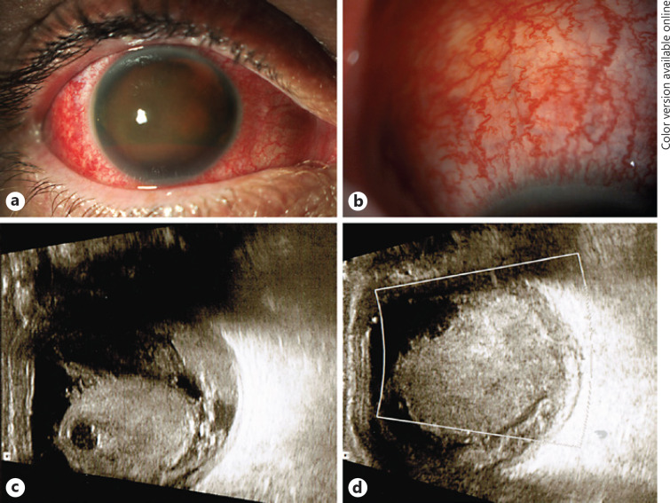
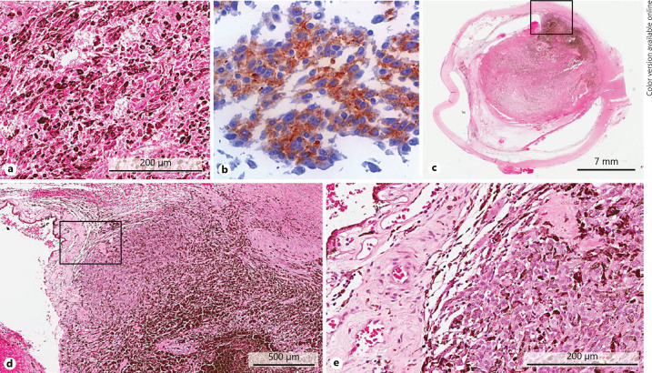
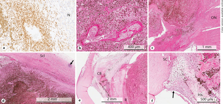
Case report: A 49-year-old man in good general health suddenly lost vision in his right eye due to an extensive vitreous haemorrhage 1 day after a workout at the gym. He reported good vision prior to that without any symptoms of flashes, floaters, or shadows. He was referred to the vitreoretinal department of a tertiary eye hospital, where he presented with a drop in vision to light perception only in the right phakic eye. Pars plana vitrectomy was performed in the right eye, which revealed intraoperatively massive retinal ischemia and choroidal haemorrhage, but no obvious tumour mass that could have been biopsied. The vitrectomy cassette specimen was sent for histopathology, where "ghost-like" melanoma cells were identified. The eye was subsequently enucleated, revealing an extensively necrotic and haemorrhagic choroidal melanoma of mixed cell type with only small viable tumour foci at the base and almost complete lysis of the detached retina.
Conclusion: Some uveal melanomas (UMs) undergo spontaneous necrosis due to rapid growth, with the centre of the tumour outstripping its established blood supply in the "watershed area" of the eye, and becoming hypoxic with associated necrosis of intraocular structures. Such UMs are often associated with haemorrhage and/or inflammation and usually cause significant destruction of ocular tissues, resulting in enucleation as the only treatment option.
Keywords: Hyphaema; Necrotic melanoma; Spontaneous necrosis; Vitreous haemorrhage.
Copyright © 2019 by S. Karger AG, Basel.
Conflict of interest statement
None of the authors has any financial relationship or conflict of interest to disclose.
Figures
Similar articles
-
Unusual presentation of a choroidal melanoma.BMJ Case Rep. 2021 May 27;14(5):e240983. doi: 10.1136/bcr-2020-240983. BMJ Case Rep. 2021. PMID: 34045197 Free PMC article.
-
DIAGNOSTIC CHALLENGES IN NECROTIC UVEAL MELANOMA.Retin Cases Brief Rep. 2022 Jan 1;16(1):92-94. doi: 10.1097/ICB.0000000000000913. Retin Cases Brief Rep. 2022. PMID: 31425450
-
Metastatic Cutaneous Melanoma Presenting with Choroidal Metastasis Mimicking Lymphoma: A Case Report.Case Rep Ophthalmol. 2025 Apr 24;16(1):360-365. doi: 10.1159/000544926. eCollection 2025 Jan-Dec. Case Rep Ophthalmol. 2025. PMID: 40463515 Free PMC article.
-
Two discrete choroidal melanomas in an eye with ocular melanocytosis.Surv Ophthalmol. 2002 Jan-Feb;47(1):36-41. doi: 10.1016/s0039-6257(01)00281-8. Surv Ophthalmol. 2002. PMID: 11801268 Review.
-
Choroidal melanoma with pigment dispersion in vitreous and melanomalytic glaucoma.Ophthalmology. 1988 Mar;95(3):370-7. doi: 10.1016/s0161-6420(88)33186-6. Ophthalmology. 1988. PMID: 3050685 Review.
Cited by
-
Description and Characteristics of Ocular Tumor Lysis Syndrome.Ocul Oncol Pathol. 2024 Sep;10(3):139-145. doi: 10.1159/000538761. Epub 2024 Apr 27. Ocul Oncol Pathol. 2024. PMID: 39224522 Free PMC article.
-
Necrosis of uveal melanoma post-COVID-19 vaccination.Indian J Ophthalmol. 2022 May;70(5):1837-1840. doi: 10.4103/ijo.IJO_3040_21. Indian J Ophthalmol. 2022. PMID: 35502089 Free PMC article.
-
Histopathologic and MR Imaging Appearance of Spontaneous and Radiation-Induced Necrosis in Uveal Melanomas: Initial Results.Cancers (Basel). 2022 Jan 2;14(1):215. doi: 10.3390/cancers14010215. Cancers (Basel). 2022. PMID: 35008378 Free PMC article.
-
Concurrence of peripunctal nevus and uveal melanoma with scleral pigment dispersion presenting as phthisis bulbi.BMJ Case Rep. 2021 Apr 16;14(4):e240854. doi: 10.1136/bcr-2020-240854. BMJ Case Rep. 2021. PMID: 33863771 Free PMC article.
References
-
- Scotto J, Fraumeni JF, Jr, Lee JA. Melanomas of the eye and other noncutaneous sites: epidemiologic aspects. J Natl Cancer Inst. 1976 Mar;56((3)):489–91. - PubMed
-
- Damato EM, Damato BE. Detection and time to treatment of uveal melanoma in the United Kingdom: an evaluation of 2,384 patients. Ophthalmology. 2012 Aug;119((8)):1582–9. - PubMed
-
- Char DH, Stone RD, Irvine AR, Crawford JB, Hilton GF, Lonn LI, et al. Diagnostic modalities in choroidal melanoma. Am J Ophthalmol. 1980 Feb;89((2)):223–30. - PubMed
Publication types
LinkOut - more resources
Full Text Sources
Miscellaneous
