. 2020 Jun 3;2(6):1414-1445.
doi: 10.1016/j.matt.2020.03.020.

Skin-interfaced sensors in digital medicine: from materials to applications

Affiliations

Skin-interfaced sensors in digital medicine: from materials to applications

Changhao Xu et al. Matter. .

Abstract

The recent advances in skin-interfaced wearable sensors have enabled tremendous potential towards personalized medicine and digital health. Compared with traditional healthcare, wearable sensors could perform continuous and non-invasive data collection from the human body and provide an insight into both fitness monitoring and medical diagnostics. In this review, we summarize the latest progress of skin-interfaced wearable sensors along with their integrated systems. We first introduce the strategies of materials selection and structure design that can be accommodated for intimate contact with human skin. Current development of physical and biochemical sensors is then classified and discussed with an emphasis on their sensing mechanisms. System-level integration including power supply, wireless communication and data analysis are also briefly discussed. We conclude with an outlook of this field and identify the key challenges and opportunities for future wearable devices and systems.

Keywords: digital health; electronic skin; flexible electronics; soft materials; wearable sensors.


Figures

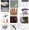
Figure 1.
Figure 1.. Soft electronic materials for skin-interfaced wearable sensors.
(A) Comparison of Young’s moduli among representative materials. Abbreviations: Nanoparticles (NP), carbon nanotubes (CNT). (B) 3D printed liquid metal arrays. (C) A twisted liquid-state heterojunction sensor based on ionic liquid. (D) A freestanding conductive hydrogel-based electrode array on soft jelly substrate. (E) Self-healing hydrogels being stretched to five times its original length. (F) A stretchable patterned PEDOT/STEC film on SEBS substrate. (G) A semiconducting P3HT based e-finger touching an ice cube. (H) SEM images of elastic conductors formed of silver nanoparticles and flakes with surfactant. (I) An AFM phase image of spray-coated carbon nanotubes on PDMS substrates. Reprinted with permission from (B) Ladd et al. Copyright 2013, WILEY-VCH Verlag GmbH & Co. KGaA, Weinheim. (C) Ota et al. Copyright 2014, Springer Nature. (D) Liu et al. Copyright 2019, Springer Nature. (E) Cao et al. Copyright 2017, WILEY-VCH Verlag GmbH & Co. KGaA, Weinheim. (F) Wang et al. Copyright 2017, AAAS. (G) Zhang et al. Copyright 2015, Springer Nature. (H) Matsuhisa et al. Copyright 2017, Springer Nature. (I) Lipomi et al. Copyright 2011, Springer Nature.
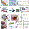
Figure 2.
Figure 2.. Structural designs for skin-interfaced wearable sensors.
(A) Paper-like ultrathin plastic electronic foils. (B) Photograph of an organic memory cells array on a plastic substrate. (C) Image of an NFC enabled pulse oximeter. (D) Controlled buckling of Si ribbons on elastomeric substrates. (E) Stretchable and foldable CMOS circuit encapsulated in wrinkled PDMS. (F) A compressed origami lithium ion battery. (G) Image of metal wires on skin-replica. (H) Image of a conductive helical coil network as interconnects for soft electronics. (I) A stretchable electronic system that integrates rigid device circuits and a serpentine interconnect network in microfluidic suspensions. (J) Image of a stretched pressure and thermal sensor network. (K) A spider-web-like conductive network mounted on a hand. (L) Schematic of microstructured Ag–Au nanocomposite in a soft matrix. Reprinted with permission from (A) Kaltenbrunner et al. Copyright 2013, Springer Nature. (B) Sekitani et al. Copyright 2009, AAAS. (C) Kim et al. Copyright 2017, WILEY-VCH Verlag GmbH & Co. KGaA, Weinheim. (D) Sun et al. Copyright 2006, Springer Nature. (E) Kim et al. Copyright 2008, AAAS. (F) Song et al. Copyright 2014, Springer Nature. (G) Fan et al. Copyright 2014, Springer Nature. (H) Jang et al. Copyright 2017, Springer Nature. (I) Xu et al. Copyright 2014, AAAS. (J) Someya et al. Copyright 2005, National Academy of Sciences. (K) Lanzara et al. Copyright 2010, WILEY-VCH Verlag GmbH & Co. KGaA, Weinheim. (L) Choi et al. Copyright 2018, Springer Nature.
Figure 3.
Figure 3.. Skin-electronics interface of wearable sensors.
(A) Tattoo-like epidermal ECG/EMG electronic system on skin. (B) A lactate tattoo sensor applied to human skin. (C) Photograph of gas-permeable nanomesh conductors attached to a fingertip. (D) A pressure sensor measuring radial artery attached to the human wrist using an adhesive bandage. (E) A flexible fully integrated wearable sensor array wristband for multiplexed in situ sweat analysis. (F) Photograph of a graphene-based glucose monitoring band on the forearm of a test person. (G) Image of a metamaterial textile sensor network for wireless communication. (H) Photograph of textile pressure sensors mounted on fingers of an artificial hand. (I) Image of a fabric-based triboelectric generator applied to a knit shirt for activity monitoring. Reprinted with permission from (A) Kim et al. Copyright 2011, AAAS. (B) Jia et al. Copyright 2013, American Chemical Society. (C) Miyamoto et al. Copyright 2017, Springer Nature. (D) Schwartz et al. Copyright 2013, Springer Nature. (E) Gao et al. Copyright 2016, Springer Nature. (F) Lipani et al. Copyright 2018, Springer Nature. (G) Tian et al. Copyright 2019, Springer Nature. (H) Liu et al. Copyright 2017, WILEY-VCH Verlag GmbH & Co. KGaA, Weinheim. (I) Jung et al. Copyright 2014, WILEY-VCH Verlag GmbH & Co. KGaA, Weinheim.
Figure 4.
Figure 4.. Temperature sensing.
(A) A core body temperature detection system based on a thermopile IR sensor mounted on ear. (B) Characterization of skin and core body temperatures as a function of environmental temperature. (C) Calibration of reflectance of thermochromic liquid crystals (TLC). (D) Optical image and infrared image of a colorimetric temperature sensor based on pixelated arrays of TLCs on the wrist. (E) Image of a single temperature sensor based on temperature coefficient of resistance (TCR). (F) Infrared image of a TCR device array mounted on the skin of the human wrist (left) and its corresponding temperature mapping (right). (G) Image of an integrated circuit for strain-independent temperature sensing. (H) Schematic of the structure of carbon nanotube (CNT) thin-film transistors (TFT) for circuits. (I) Demonstration of stable functionality of temperature sensor attached to a prosthetic hand during repeated bending. Reprinted with permission from (A,B) Ota et al. Copyright 2017, American Chemical Society. (C,D) Gao et al. Copyright 2014, Springer Nature. (E,F) Webb et al. Copyright 2013, Springer Nature. (G-I) Zhu et al. Copyright 2018, Springer Nature.
Figure 5.
Figure 5.. Motion and tactile sensing.
(A) A CNT strain sensor array fixed to a data glove. (B) SEM image of the fractural structure of the SWCNT film at 100% strain. (C) Relative changes in resistance versus time for data glove motion of gestures. (D) Cross sectional HRTEM image of a single pressure-sensitive nanofibre. (E) Performance of the pressure response of the device in various bending states. (F,G) Current mapping of a stretchable transistor array, matching exactly with the position of a ladybug. (H) Schematic of a single pixel of the user-interactive e-skin. (I) Image of interactive e-skin device showing local light emission where the surface is touched. (J) Exploded view of the artificial skin of a prosthetic hand. (K) An image of the prosthetic limb catching a baseball, and the corresponding signal of the tactile sensor. Reprinted with permission from (A–C) Yamada et al. Copyright 2011, Springer Nature. (D,E) Lee et al. Copyright 2016, Springer Nature. (F,G) Wang et al. Copyright 2018, Springer Nature. (H,I) Wang et al. Copyright 2013, Springer Nature. (J,K) Kim et al. Copyright 2014, Springer Nature.
Figure 6.
Figure 6.. Vascular dynamics monitoring.
(A) Photograph of a flexible resistive sensor for wrist pulse detection. (B) Schematic of the pulse sensing e-skin. (C) Signals measuring wrist pulses of a healthy person and a pregnant woman. (D) Schematic of a pressure sensor with microhair structures. (E) Pulse waves of the radial artery measured with PDMS microhair sensors. (F) A moisture-impermeable ultrasonic device conforming to complex surfaces. (G) Sensing mechanism schematic of the pulse-echo method using an ultrasonic beam. (H) Pulse waveform comparison measured before and after exercise. (I) Photograph of an organic optical sensor mounted on a finger. (J) Device structure and operation principle of the reflective pulse oximeter. (K) Output signal from OPD with varying oxygenation of blood. Reprinted with permission from (A-C) Wang et al. Copyright 2013, WILEY-VCH Verlag GmbH & Co. KGaA, Weinheim. (D,E) Pang et al. Copyright 2015, WILEY-VCH Verlag GmbH & Co. KGaA, Weinheim. (F-H) Wang et al. Copyright 2018, Springer Nature. (I-K) Yokota et al. Copyright 2016, AAAS.
Figure 7.
Figure 7.. Electrophysiology monitoring.
(A) Image of an epidermal organic differential amplifier for electrocardiogram (ECG) monitoring. (B) Cross-sectional schematic of organic TFT and thin-film capacitor in the circuit. (C) Illustrations of ECG signal amplification and noise reduction based on differential amplifier. (D) Image of an electrophysiological wearable electronic patch. (E) Impedance comparison of the Ag–Au nanocomposite electrode and Ag/AgCl gel electrodes at the skin-electrode interface. (F) ECG and electromyogram (EMG) recordings using the wearable device on the skin. (G) Exploded view of the large-area epidermal electrodes for electroencephalogram (EEG) mapping. (H) Image comparison of epidermal (E1 and E2) and conventional EEG cup (Pz) electrodes on the scalp of a subject, and (I) corresponding recorded EEG signals. Reprinted with permission from (A-C) Sugiyama et al. Copyright 2019, Springer Nature. (D-F) Choi et al. Copyright 2018, Springer Nature. (G–I) Tian et al. Copyright 2019, Springer Nature.
Figure 8.
Figure 8.. Sweat analysis.
(A) Fully integrated headband and wristband for on-body real-time perspiration analysis during exercise. (B) Schematic of the sensor array for multi-analyte sensing. (C) A laser-engraved sensor patch for sweat uric acid (UA) and tyrosine detection. (D) Sweat and serum UA before and after purine intake on a healthy subject, measured by the laser-engraved sensor patch. (E) Image of a graphene-hybrid electrochemical patch on the human skin. (F) Schematic of the diabetes monitoring and therapy system. (G) Sweat and blood glucose concentrations on a human subject over a day. (H) Schematic of an iontophoretic device for alcohol sensing. (I) Photograph of the integrated tattoo device applied to a human subject. (K) Pharmacokinetic curve of blood and sweat alcohol concentration. (L) Schematic of colorimetric sensor patch for sweat lactate, glucose, creatinine, pH, and chloride ions determination. (M) Sweat analysis during a cycling exercise. (N) Photograph of a fluorometric sensor patch for sweat chloride, sodium, and zinc detection. (O) Calibration plot of fluorescence intensity over chloride concentrations. Reprinted with permission from (A,B) Gao et al. Copyright 2016, Springer Nature. (C,D) Yang et al. Copyright 2020, Springer Nature. (E–G) Lee et al. Copyright 2016, Springer Nature. (H,I) Kim et al. Copyright 2016, American Chemical Society. (J,K) Hauke et al. Copyright 2018, The Royal Society of Chemistry. (L,M) Koh et al. Copyright 2016, AAAS. (N,O) Sekine et al. Copyright 2018, The Royal Society of Chemistry.
Figure 9.
Figure 9.. Interstitial fluid (ISF) analysis.
(A) Schematic of the tattoo sensor for noninvasive glucose sensing based on reverse iontophoresis (RI). (B) Photograph of the tattoo-like sensing device on a human forearm. (C) Amperograms of the tattoo-glucose sensor obtained pre- and post-meal from ISF. (D) ISF sensing mechanism by using RI with electrochemical twin channels (ETC). (E) Image of RI electrodes attached to skin. (F) Hourly glucose monitoring in comparison with a glucometer. (G) Working principle of a glucose sensor array targeting transdermal individual preferential glucose pathways. (H) Photograph of a screen-printed sensor array fixed onto a subject’s forearm. (I) In vivo continuous glucose monitoring on a healthy human subject using blood glucose as a reference. (J) Demonstrations of a dual iontophoresis sensor for simultaneous sweat alcohol and ISF glucose detection. (K) Schematic of electrode layout and composition of the working electrodes. Reprinted with permission from (A–C) Bandodkar et al. Copyright 2014, American Chemical Society. (D–F) Chen et al. Copyright 2017, AAAS. (G–I) Lipani et al. Copyright 2018, Springer Nature. (J,K) Kim et al. Copyright 2018, The Authors.
Figure 10.
Figure 10.. Power supply.
(A) Exploded view of a flexible lithium-ion battery. (B) Image of the stretched battery mounted on the human elbow. (C) Photograph of a flexible organic photovoltaics device wrapped over a rod. (D) Schematic of the organic photovoltaics. (E) Schematic of an e-skin based biofuel cell and electrochemical reactions on electrodes. (F) Demonstration of the BFC worn on a human subject. (G) Demonstration of a triboelectric nanogenerator (TENG) assembled into clothes and shoes for power collection. (H) Schematic of the tube-shaped TENG device. (I) Photograph and schematic of a wearable thermoelectric device. (J) Power generation of the thermoelectric device under various thermal conditions. (K) Schematic of fiber-shaped TENG unit (top) and dye-sensitized solar cells (bottom). (L) Demonstration of the hybridized power textile during outdoor activities on a human subject. Reprinted with permission from (A,B) Xu et al. Copyright 2013, Springer Nature. (C,D) Park et al. Copyright 2018, Springer Nature. (E,F) Bandodkar et al. Copyright 2017, The Royal Society of Chemistry. (G,H) Wang et al. Copyright 2016, Springer Nature. (I,J) Hong et al. Copyright 2019, AAAS. (K,L) Wen et al. Copyright 2016, AAAS.
Figure 11.
Figure 11.. Communication and data analysis.
(A) Photograph of sweat collection at the neck of a subject under high levels of deformation. (B) Structure of NFC electronics for digital thermography and chloride sensing. (C) Image of a battery-free NFC electronic sensor patch. (D) Illustration of a phone interface for wireless communication and image acquisition. (E) Image of a Bluetooth-enabled wearable platform with electrochemical sensors connected to a flexible PCB module. (F) Demonstration of wireless sweat analysis by uploading data to cloud servers. (G) Image of a wireless sensor network composed of stretchable on-skin sensors and flexible PCB initiator. (H) Design of the stretchable sensor target node. (I) Demonstration of pulse monitoring through a sensor node placed on a subject’s wrist. Reprinted with permission from (A,B) Reeder et al. Copyright 2019, AAAS. (C,D) Bandodkar et al. Copyright 2019, AAAS. (E,F) Gao et al. Copyright 2016, Springer Nature. (G-I) Niu et al. Copyright 2019, Springer Nature.

References

    1. Ray TR, Choi J, Bandodkar AJ, Krishnan S, Gutruf P, Tian L, Ghaffari R, and Rogers JA (2019). Bio-Integrated Wearable Systems: A Comprehensive Review. Chem. Rev 119, 5461–5533. - PubMed
    1. Wang C, Wang C, Huang Z, and Xu S (2018). Materials and Structures toward Soft Electronics. Adv. Mater 30, 1801368. - PubMed
    1. Someya T, Bao Z, and Malliaras GG (2016). The rise of plastic bioelectronics. Nature 540, 379–385. - PubMed
    1. Kim J, Campbell AS, Ávila B.E.-F. de, and Wang J (2019). Wearable biosensors for healthcare monitoring. Nat. Biotechnol 37, 389–406. - PMC - PubMed
    1. Yang Y, and Gao W (2019). Wearable and flexible electronics for continuous molecular monitoring. Chem. Soc. Rev 48, 1465–1491. - PubMed

LinkOut - more resources