Case Reports
. 2020 Aug;16(2):274-277.
doi: 10.5152/iao.2019.4002.

An atypical stapedial artery

Affiliations
Case Reports

An atypical stapedial artery

Mélanie Sanjuan et al. J Int Adv Otol. 2020 Aug.

Abstract

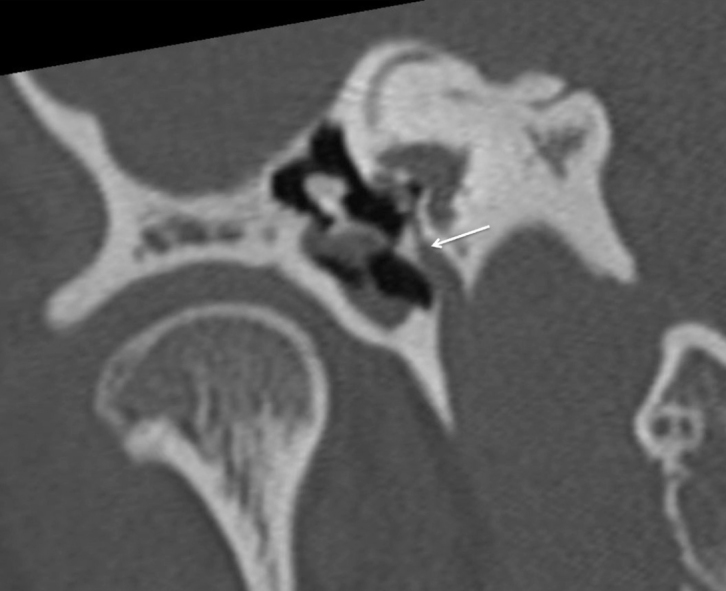
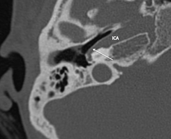
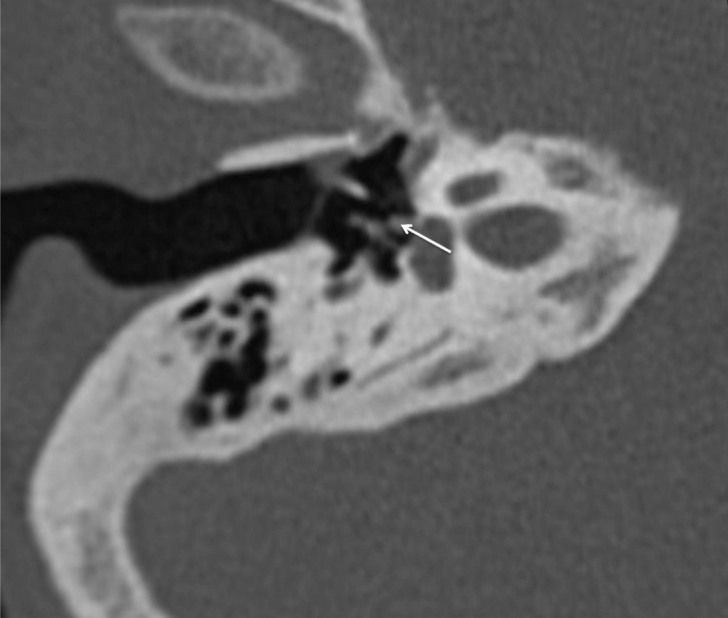
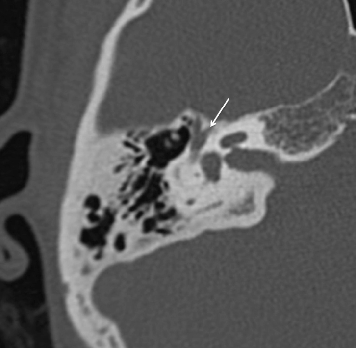
The persistence of the stapedial artery is a rare vascular anomaly. It is mostly asymptomatic but sometimes cause conductive hearing loss, pulsatile tinnitus, or vertigo. The estimated prevalence of this rare postembryonic persistence ranged from 0.02% to 0.48%. Four different anatomical forms have been identified, and their preoperative diagnostic is essential. We report the case of an incidental discovery of pharyngo-hyo-stapedial artery, the most uncommon form of persistent stapedial artery. Its per-operative finding has become rare because tomodensitometry is performed in case of conductive hearing loss. The continuous improvement of imagery resolution will probably help to revise the incidence of this malformation.


Conflict of interest statement

Conflict of Interest: The authors have no conflict of interest to declare.

Figures

Figure 1
Figure 1
Sagittal CT scan of right temporal bone showing an enlarged canal of the Jacobson’s nerve, ascending through a bony canal over the cochlear promontory (white arrow).
Figure 2
Figure 2
Axial CT of the right temporal bone showing an artery (white arrow) arising from the junction between the vertical and horizontal segments of the intra-petrous internal carotid artery.
Figure 3
Figure 3
Axial CT of the right temporal bone at the level of the stapes showing a persistent stapedial artery (white arrow).
Figure 4
Figure 4
Axial CT scan of the right temporal bone at the level of the geniculate ganglion showing the exit of the persistent stapedial artery in the middle skull base (white arrow).
Figure 5. a–f
Figure 5. a–f
Schematic representation of the embryology of the stapedial artery at seven weeks (a), its normal evolution (b), and the four anatomic variations (c, d, e, f).
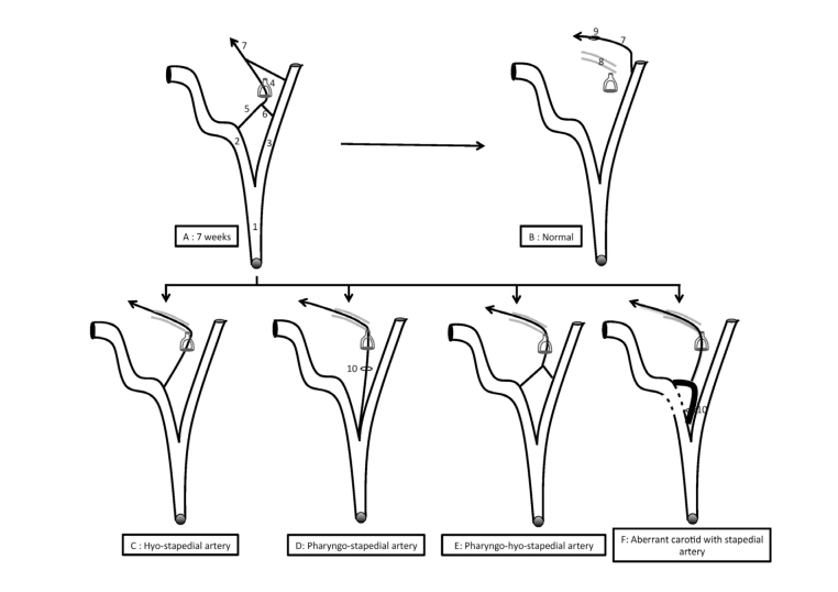
  1. Common carotid artery

  2. Internal carotid artery

  3. External carotid artery

  4. Stapes

  5. Hyoid artery

  6. Pharyngeal artery

  7. Middle meningeal artery

  8. 2° portion of the fallopian canal

  9. Foramen spinosum

  10. Tympanic canaliculus

References

    1. Schuknecht HF, Gulya AJ. Anatomy of the temporal bone with surgical implications. Philadelphia: Lea and Febiger; 1995.
    1. Hitier M, Zhang M, Labrousse M, Barbier C, Patron V, Moreau S. Persistent stapedial arteries in human: from phylogeny to surgical consequences. Surg Radiol Anat. 2013;35:883–91. doi: 10.1007/s00276-013-1127-z. - DOI - PubMed
    1. Govaerts PJ, Marquet TF, Cremers C, Offeciers FE. Persistent stapedial artery: does it prevent successful surgery? Ann Otol Rhinol Laryngol. 1993;102:724–8. doi: 10.1177/000348949310200914. - DOI - PubMed
    1. Moreano EH, Puparella MM, Zelterman D, Goycoolea MV. Prevalence of facial canal dehiscence and of persistent stapedial artery in the human middle ear: a report of 1000 temporal bones. Laryngoscope. 1994;104:309–20. doi: 10.1288/00005537-199403000-00012. - DOI - PubMed
    1. Altman F. Anomalies of the internal carotid artery and its branches; their embryological and comparative anatomical significance: report of a new case of persistent stapedial artery in man. Laryngoscope. 1947;57:313–39. doi: 10.1288/00005537-194705000-00002. - DOI - PubMed

Publication types