[Preprint]. 2020 Feb 21.
doi: 10.26434/chemrxiv.11875446.

Fast Identification of Possible Drug Treatment of Coronavirus Disease -19 (COVID-19) Through Computational Drug Repurposing Study

Affiliations

Fast Identification of Possible Drug Treatment of Coronavirus Disease -19 (COVID-19) Through Computational Drug Repurposing Study

Junmei Wang. ChemRxiv. .

Update in

Abstract

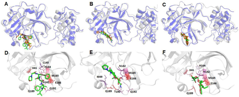
The recent outbreak of novel coronavirus disease -19 (COVID-19) calls for and welcomes possible treatment strategies using drugs on the market. It is very efficient to apply computer-aided drug design techniques to quickly identify promising drug repurposing candidates, especially after the detailed 3D-structures of key virous proteins are resolved. Taking the advantage of a recently released crystal structure of COVID-19 protease in complex with a covalently-bonded inhibitor, N3,1 I conducted virtual docking screening of approved drugs and drug candidates in clinical trials. For the top docking hits, I then performed molecular dynamics simulations followed by binding free energy calculations using an endpoint method called MM-PBSA-WSAS.2-4 Several promising known drugs stand out as potential inhibitors of COVID-19 protease, including Carfilzomib, Eravacycline, Valrubicin, Lopinavir and Elbasvir. Carfilzomib, an approved anti-cancer drug acting as a proteasome inhibitor, has the best MM-PBSA-WSAS binding free energy, -13.82 kcal/mol. Streptomycin, an antibiotic and a charged molecule, also demonstrates some inhibitory effect, even though the predicted binding free energy of the charged form (-3.82 kcal/mol) is not nearly as low as that of the neutral form (-7.92 kcal/mol). One bioactive, PubChem 23727975, has a binding free energy of -12.86 kcal/mol. Detailed receptor-ligand interactions were analyzed and hot spots for the receptor-ligand binding were identified. I found that one hotspot residue HIS41, is a conserved residue across many viruses including COVID-19, SARS, MERS, and HCV. The findings of this study can facilitate rational drug design targeting the COVID-19 protease.


Conflict of interest statement

The author declares no competing financial interest.

Figures

Figure 1.
Figure 1.
2D-Structures of promising repurpose drugs. All five approved drugs are in neural form under physiological conditions.
Figure 2.
Figure 2.
2D-Structures of promising repurposing drugs. All three approved drugs are in charged form under physiological conditions.
Figure 3.
Figure 3.
2D-Structures of promising bio-actives which are structurally similar to Lopinavir. PubChem 88143175, although studied in neutral form, bears −3 charges under physiological conditions.
Figure 4.
Figure 4.
Plots of Root-mean-square deviations of receptor main chain atoms and ligand heavy atoms along the MD simulation time for (A) co-crystal ligand N3, (B) DB08889, (C) DB12329, (D) DB00385, (E) DB01601 and (F) DB11574.
Figure 5.
Figure 5.
Structural comparison between the crystal structure and a representative MD structureof COVID-19 protease bound to the known ligand, N3 . The crystal structure is shown as blue cartoon with the co-crystal ligand shown as brown sticks, while the representative MD structure is shown in grey cartoon and the ligand as green sticks (Panel A). The hotspot residues (ΔGLig-Res < −3.0 kcal/mol) revealed by MM-GBSA analysis are shown in Panel B; the more bluish a residue is colored, the stronger the interaction between the residue and the ligand.
Figure 6.
Figure 6.
Structural comparison between the crystal structure and a representative MD structure of COVID-19 protease bound to three neutral ligands DB08889, DB12329, and DB00385. The crystal structure is shown as blue cartoon with the docked ligand shown as brown sticks, while the representative MD structure is shown in grey cartoon and the ligand as green sticks. A: DB08889, B: DB12329, and C: DB00385. The detailed ligand-receptor interactions are shown in the bottom panel (D-F). All the hotspot residues (ΔGLig-Res < −3.0) revealed by MM-GBSA analyses are labeled and colored by a blue to red spectrum, the more bluish a residue is colored, the stronger the interaction between the residue and the ligand. D: DB08889, E: DB12329, and F: DB00385.
Figure 7.
Figure 7.
Structural comparison between the crystal and a representative MD structure of COVID-19 protease bound to two neutral ligands DB01601 and DB11574. The crystal structure is shown as blue cartoon with the docked ligand shown as brown sticks, while the representative MD structure is shown in grey cartoon and the ligand as green sticks. A: DB01601 and B: DB11574. The detailed ligand-receptor interactions are shown in the bottom panel (C-D). All the hotspot residues (ΔGLig-Res < −3.0) revealed by MM-GBSA analyses are labeled and colored by a blue to red spectrum, the more bluish a residue is colored, the stronger interaction between the residue and the ligand. C: DB01601 and D: DB11574.
Figure 8.
Figure 8.
Structural comparison between the crystal structure and a representative MD structure of COVID-19 protease bound to three charged ligands DB01082, DB03147, and DB11184. The crystal structure is shown as blue cartoon with the docked ligand shown as brown sticks, while the representative MD structure is shown in grey cartoon and the ligand as green sticks. A: DB01082, B: DB03147, and C: DB11184. The detailed ligand-receptor interactions are shown in the bottom panel (D-F). All the hotspot residues (ΔGLig-Res < −3.0) revealed by MM-GBSA analyses are labeled and colored by a blue to red spectrum, the more bluish a residue is colored, the stronger the interaction between the residue and the ligand. D: DB01082, E: DB03147, and F: DB11184.
Figure 9.
Figure 9.
Structural comparison of proteases among three coronavirus viruses (COVID-19, SARS and MERS) (A), and between COVID-19 and hepatitis C NS3/4A proteases (B). The COVID-19 protease is colored in grey and its ligands are shown as green sticks. The following are the color codes for the other proteases: SARS protease and its co-crystal ligand - brown, MERS protease and its co-crystal ligand - blue, HCV NS3/4A - blue, co-crystal ligand of HCV NS3/4A - brown. Backbone RMSD between SARS and COVID-19 is 0.4711 Å, with 284 residues participating in the least-square fitting and 22 omitted, and the backbone RMSD between MERS and COVID-19 is 0.41 Å, but with 195 residues participating in the least-square fitting and 104 omitted. In contrast, the backbone RMSD between COVID-19 protease and HCV NS3/4A is 2.2632 Å, with 108 residues participating in the least-square fitting and 43 omitted.

References

    1. Liu X.; Zhang B.; Jin Z.; Yang H.; Rao Z., The crystal structure of COVID-19 main protease in complex with an inhibitor N3. 2020.
    1. Wang E.; Sun H.; Wang J.; Wang Z.; Liu H.; Zhang J. Z. H.; Hou T., End-Point Binding Free Energy Calculation with MM/PBSA and MM/GBSA: Strategies and Applications in Drug Design. Chem Rev 2019, 119 (16), 9478–9508. - PubMed
    1. Wang J. M.; Hou T. J.; Xu X. J., Recent Advances in Free Energy Calculations with a Combination of Molecular Mechanics and Continuum Models. Curr Comput-Aid Drug 2006, 2 (3), 287–306.
    1. Wang J.; Hou T., Develop and test a solvent accessible surface area-based model in conformational entropy calculations. Journal of chemical information and modeling 2012, 52 (5), 1199–212. - PMC - PubMed
    1. Law V.; Knox C.; Djoumbou Y.; Jewison T.; Guo A. C.; Liu Y.; Maciejewski A.; Arndt D.; Wilson M.; Neveu V.; Tang A.; Gabriel G.; Ly C.; Adamjee S.; Dame Z. T.; Han B.; Zhou Y.; Wishart D. S., DrugBank 4.0: shedding new light on drug metabolism. Nucleic acids research 2014, 42 (Database issue), D1091–7. - PMC - PubMed

Publication types

LinkOut - more resources