

Single-cell RNA analysis on ACE2 expression provides insights into SARS-CoV-2 potential entry into the bloodstream and heart injury
- PMID: 32510598
- PMCID: PMC7301022
- DOI: 10.1002/jcp.29802
Single-cell RNA analysis on ACE2 expression provides insights into SARS-CoV-2 potential entry into the bloodstream and heart injury
Abstract
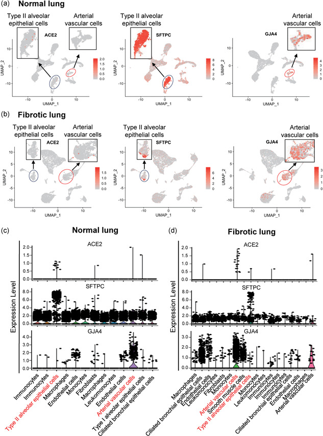
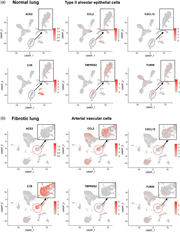
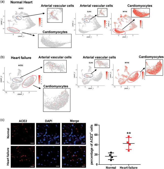
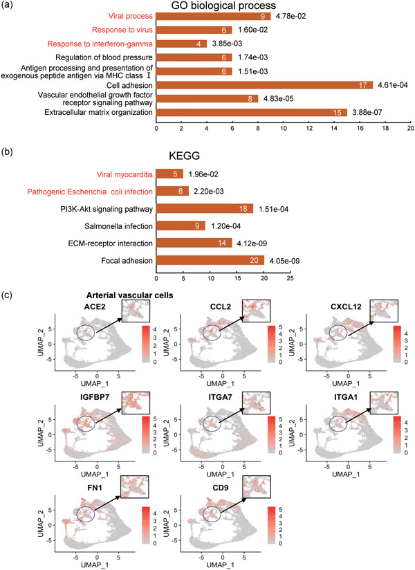
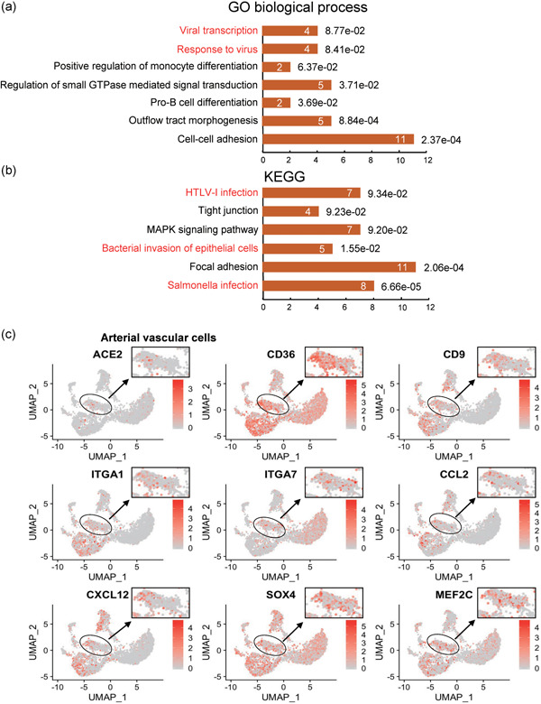
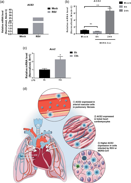
Coronavirus disease-2019 (COVID-19) is a global pandemic with high infectivity and pathogenicity, accounting for tens of thousands of deaths worldwide. Recent studies have found that the pathogen of COVID-19, severe acute respiratory syndrome coronavirus 2 (SARS-CoV-2), shares the same cell receptor angiotensin converting enzyme II (ACE2) as SARS-CoV. The pathological investigation of COVID-19 deaths showed that the lungs had characteristics of pulmonary fibrosis. However, how SARS-CoV-2 spreads from the lungs to other organs has not yet been determined. Here, we performed an unbiased evaluation of cell-type-specific expression of ACE2 in healthy and fibrotic lungs, as well as in normal and failed adult human hearts, using published single-cell RNA-seq data. We found that ACE2 expression in fibrotic lungs mainly locates in arterial vascular cells, which might provide a route for bloodstream spreading of SARS-CoV-2. Failed human hearts have a higher percentage of ACE2-expressing cardiomyocytes, and SARS-CoV-2 might attack cardiomyocytes through the bloodstream in patients with heart failure. Moreover, ACE2 was highly expressed in cells infected by respiratory syncytial virus or Middle East respiratory syndrome coronavirus and in mice treated by lipopolysaccharide. Our findings indicate that patients with pulmonary fibrosis, heart failure, and virus infection have a higher risk and are more susceptible to SARS-CoV-2 infection. The SARS-CoV-2 might attack other organs by getting into the bloodstream. This study provides new insights into SARS-CoV-2 blood entry and heart injury and might propose a therapeutic strategy to prevent patients from developing severe complications.
Keywords: ACE2; COVID-19; SARS-CoV2; heart failure; pulmonary fibrosis.
© 2020 Wiley Periodicals LLC.
Conflict of interest statement
The authors declare that there are no conflicts of interest.
Figures
Similar articles
-
Single-cell analysis of SARS-CoV-2 receptor ACE2 and spike protein priming expression of proteases in the human heart.Cardiovasc Res. 2020 Aug 1;116(10):1733-1741. doi: 10.1093/cvr/cvaa191. Cardiovasc Res. 2020. PMID: 32638018 Free PMC article.
-
Expressions and significances of the angiotensin-converting enzyme 2 gene, the receptor of SARS-CoV-2 for COVID-19.Mol Biol Rep. 2020 Jun;47(6):4383-4392. doi: 10.1007/s11033-020-05478-4. Epub 2020 May 14. Mol Biol Rep. 2020. PMID: 32410141 Free PMC article.
-
SARS-CoV-2 strategically mimics proteolytic activation of human ENaC.Elife. 2020 May 26;9:e58603. doi: 10.7554/eLife.58603. Elife. 2020. PMID: 32452762 Free PMC article.
-
Role of angiotensin-converting enzyme 2 and pericytes in cardiac complications of COVID-19 infection.Am J Physiol Heart Circ Physiol. 2020 Nov 1;319(5):H1059-H1068. doi: 10.1152/ajpheart.00681.2020. Epub 2020 Oct 9. Am J Physiol Heart Circ Physiol. 2020. PMID: 33036546 Free PMC article. Review.
-
SARS-CoV-2, More than a Respiratory Virus: Its Potential Role in Neuropathogenesis.ACS Chem Neurosci. 2020 Jul 1;11(13):1887-1899. doi: 10.1021/acschemneuro.0c00251. Epub 2020 Jun 18. ACS Chem Neurosci. 2020. PMID: 32491829 Review.
Cited by
-
Investigating the ACE2 polymorphisms in COVID-19 susceptibility: An in silico analysis.Mol Genet Genomic Med. 2021 Jun;9(6):e1672. doi: 10.1002/mgg3.1672. Epub 2021 Apr 5. Mol Genet Genomic Med. 2021. PMID: 33818000 Free PMC article.
-
Brazilian Society of Cardiology Guideline on Myocarditis - 2022.Arq Bras Cardiol. 2022 Jul;119(1):143-211. doi: 10.36660/abc.20220412. Arq Bras Cardiol. 2022. PMID: 35830116 Free PMC article. English, Portuguese. No abstract available.
-
SARS-CoV-2 cellular tropism and direct multiorgan failure in COVID-19 patients: Bioinformatic predictions, experimental observations, and open questions.Cell Biol Int. 2023 Feb;47(2):308-326. doi: 10.1002/cbin.11928. Epub 2022 Oct 13. Cell Biol Int. 2023. PMID: 36229927 Free PMC article. Review.
-
Angiotensin-converting enzyme 2: a double-edged sword in COVID-19 patients with an increased risk of heart failure.Heart Fail Rev. 2021 Mar;26(2):371-380. doi: 10.1007/s10741-020-10016-2. Heart Fail Rev. 2021. PMID: 32844337 Free PMC article. Review.
-
Innate Immunity Plays a Key Role in Controlling Viral Load in COVID-19: Mechanistic Insights from a Whole-Body Infection Dynamics Model.ACS Pharmacol Transl Sci. 2020 Dec 30;4(1):248-265. doi: 10.1021/acsptsci.0c00183. eCollection 2021 Feb 12. ACS Pharmacol Transl Sci. 2020. PMID: 33615177 Free PMC article.
References
-
- Hamming, I. , Timens, W. , Bulthuis, M. L. , Lely, A. T. , Navis, G. , & van Goor, H. (2004). Tissue distribution of ACE2 protein, the functional receptor for SARS coronavirus. A first step in understanding SARS pathogenesis. Journal of Pathology, 203(2), 631–637. 10.1002/path.1570 - DOI - PMC - PubMed
MeSH terms
Substances
Grants and funding
- 81873536/General Programs of the National Natural Science Foundation of China/International
- 81572713/General Programs of the National Natural Science Foundation of China/International
- 81873469 and 81670450/General Programs of the National Natural Science Foundation of China/International
- 2019M651371/China Postdoctoral Science Foundation/International
- 2019T120303 and 2019T120303/China Postdoctoral Science Foundation/International
LinkOut - more resources
Full Text Sources
Miscellaneous
