Review
. 2020 Aug;40(8):1818-1829.
doi: 10.1161/ATVBAHA.120.314558. Epub 2020 Jun 8.

Overcoming Barriers: The Endothelium As a Linchpin of Coronavirus Disease 2019 Pathogenesis?

Affiliations
Review

Overcoming Barriers: The Endothelium As a Linchpin of Coronavirus Disease 2019 Pathogenesis?

Dakota Gustafson et al. Arterioscler Thromb Vasc Biol. 2020 Aug.

Abstract

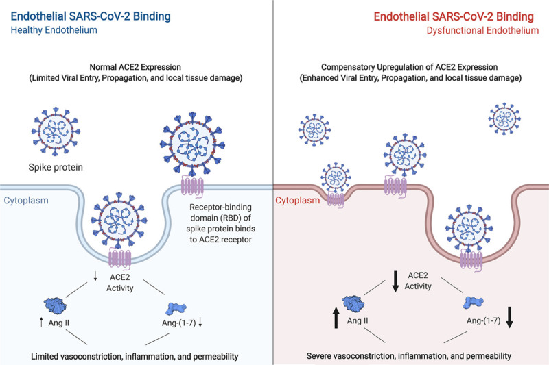
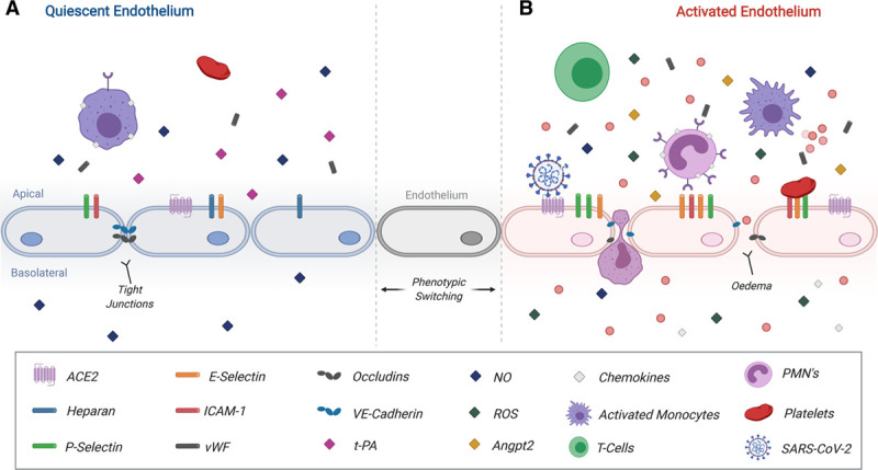
Objective: Coronavirus disease 2019 (COVID-19) is a global pandemic involving >5 500 000 cases worldwide as of May 26, 2020. The culprit is the severe acute respiratory syndrome coronavirus-2, which invades cells by binding to ACE2 (angiotensin-converting enzyme 2). While the majority of patients mount an appropriate antiviral response and recover at home, others progress to respiratory distress requiring hospital admission for supplemental oxygen. In severe cases, deterioration to acute respiratory distress syndrome necessitating mechanical ventilation, development of severe thrombotic events, or cardiac injury and dysfunction occurs. In this review, we highlight what is known to date about COVID-19 and cardiovascular risk, focusing in on the putative role of the endothelium in disease susceptibility and pathogenesis. Approach and Results: Cytokine-driven vascular leak in the lung alveolar-endothelial interface facilitates acute lung injury in the setting of viral infection. Given that the virus affects multiple organs, including the heart, it likely gains access into systemic circulation by infecting or passing from the respiratory epithelium to the endothelium for viral dissemination. Indeed, cardiovascular complications of COVID-19 are highly prevalent and include acute cardiac injury, myocarditis, and a hypercoagulable state, all of which may be influenced by altered endothelial function. Notably, the disease course is worse in individuals with preexisting comorbidities that involve endothelial dysfunction and may be linked to elevated ACE2 expression, such as diabetes mellitus, hypertension, and cardiovascular disease.

Conclusions: Rapidly emerging data on COVID-19, together with results from studies on severe acute respiratory syndrome coronavirus-1, are providing insight into how endothelial dysfunction may contribute to the pandemic that is paralyzing the globe. This may, in turn, inform the design of biomarkers predictive of disease course, as well as therapeutics targeting pathogenic endothelial responses.

Keywords: biomarkers; cardiac injury; coronavirus; endothelium; pandemic.


Figures

Figure 1.
Figure 1.
Proposed model of the pathological effect of severe acute respiratory syndrome coronavirus-2 (SARS-CoV-2) on the endothelium. Left, under conditions of endothelial quiescence, binding of SARS-CoV-2 is mediated in part by ACE2 (angiotensin-converting enzyme 2). Internalization of ACE2, and subsequent receptor interference, results in an upregulation of AngII (angiotensin II) and downregulation of Ang-(1-7) (angiotensin-[1-7]). Although contributing to vasoconstriction, inflammation, and permeability, limited expression of ACE2 within a relatively quiescent endothelium may result in limited viral entry and local and systemic dysfunction. Right, paradoxically, although a dysfunctional endothelium may have higher baseline levels of ACE2, enhanced viral entry may increase the degradation of ACE2 in the lysosome, enhancing inflammation and tissue damage. The resulting downregulation of ACE2 activity, upregulation of Ang-II, and downregulation of Ang-(1-7) thereby may have a starker induction of vasoconstriction, inflammation, and permeability as compensatory mechanisms are abrogated. Arrow width corresponds to intensity. This figure was created with the assistance of www.BioRender.com.
Figure 2.
Figure 2.
Proposed pathobiology of activated endothelium during severe acute respiratory syndrome coronavirus-2 (SARS-CoV-2 infection). A, A quiescent endothelium is shown on the left. Endothelial quiescence facilitates normal responses to angiogenic signalling, facilitates controlled immune surveillance, and provides homeostatic cues for coagulation. B, Chronic activation of the endothelium is marked by stark upregulation of P-selectin, E-Selectin, ICAM-1, and ACE2 (angiotensin-converting enzyme 2), along with their respective soluble forms. The subsequent loss of tight junction expression expedites edema and facilitates enhanced recruitment, attachment, and extravasation of immune cells across the vascular endothelium. Disruption of coagulation cues through the loss of heparan, DDP-4, and t-PA (tissue-type plasminogen activator) and secretion of vWF (von Willebrand Factor) exacerbates endothelial injury and induces coagulopathies. Further disruption of endothelial phenotype results in local reductions of secreted NO, increases in secreted reactive oxygen species (ROS), and enhanced Angpt2 secretion. SARS-CoV-2 reduces ACE2 availability further propagating endothelial dysfunction. While intended to assist in infection control, innate and adaptive immune responses can instead induce a storm of chemokines and cytokines, which further propagates underlying inflammation and dysfunction. This figure was created with the assistance of www.BioRender.com. ICAM-1 indicates intercellular adhesion molecule; NO, nitric oxide; PMN, polymorphonuclear leukocyte; and VE-Cadherin, vascular endothelial cadherin.

References

    1. Lu R, Zhao X, Li J, Niu P, Yang B, Wu H, Wang W, Song H, Huang B, Zhu N, et al. Genomic characterisation and epidemiology of 2019 novel coronavirus: implications for virus origins and receptor binding. Lancet 2020395565–574doi: 10.1016/S0140-6736(20)30251-8 - PMC - PubMed
    1. World Health Organization. Novel Coronavirus (2019-nCoV)-SITUATION REPORT-1. 2020 https://www.who.int/docs/default-source/coronaviruse/situation-reports/2.... Accessed April 6, 2020.
    1. Li LQ, Huang T, Wang YQ, Wang Z-P, Liang Y, Huang T-B, Zhang H-Y, Sun W, Wang Y. COVID-19 patients’ clinical characteristics, discharge rate, and fatality rate of meta-analysis [published online March 12, 2020 ]. J Med Virol. doi: 10.1002/jmv.25757. - PMC - PubMed
    1. Rodriguez-Morales AJ, Cardona-Ospina JA, Gutiérrez-Ocampo E, Villamizar-Peña R, Holguin-Rivera Y, Escalera-Antezana JP, Alvarado-Arnez LE, Bonilla-Aldana DK, Franco-Paredes C, Henao-Martinez AF, et al. ; Latin American Network of Coronavirus Disease 2019-COVID-19 Research (LANCOVID-19) Electronic address: https://www.lancovid.org. Clinical, laboratory and imaging features of COVID-19: A systematic review and meta-analysis. Travel Med Infect Dis 202034101623.doi: 10.1016/j.tmaid.2020.101623 - PMC - PubMed
    1. Grasselli G, Zangrillo A, Zanella A, Antonelli M, Cabrini L, Castelli A, Cereda D, Coluccello A, Foti G, Fumagalli R, et al. Baseline characteristics and outcomes of 1591 patients infected with SARS-CoV-2 admitted to ICUs of the Lombardy Region, Italy. JAMA 20203231574–1581 - PMC - PubMed

Publication types

MeSH terms