. 2020 Jun 8;16(6):e1008803.
doi: 10.1371/journal.pgen.1008803. eCollection 2020 Jun.

MYO5B mutations in pheochromocytoma/paraganglioma promote cancer progression

Affiliations

MYO5B mutations in pheochromocytoma/paraganglioma promote cancer progression

Tajana Tešan Tomić et al. PLoS Genet. .

Abstract

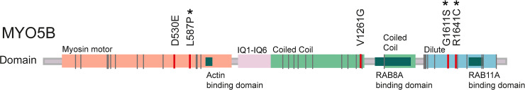
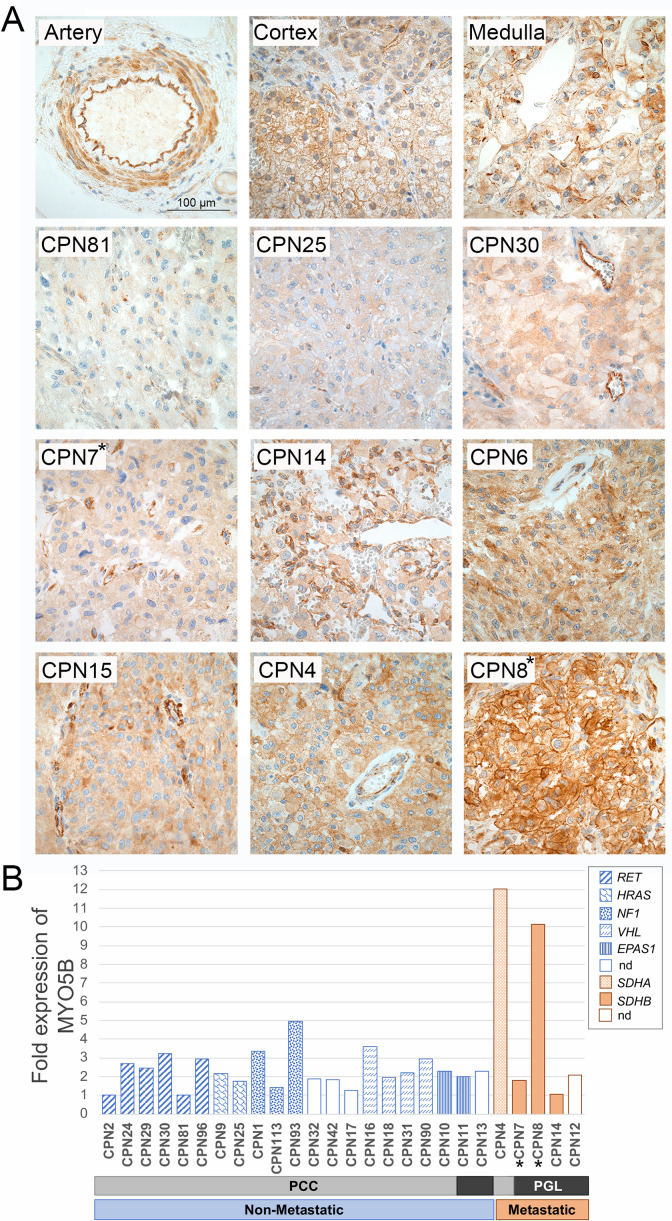
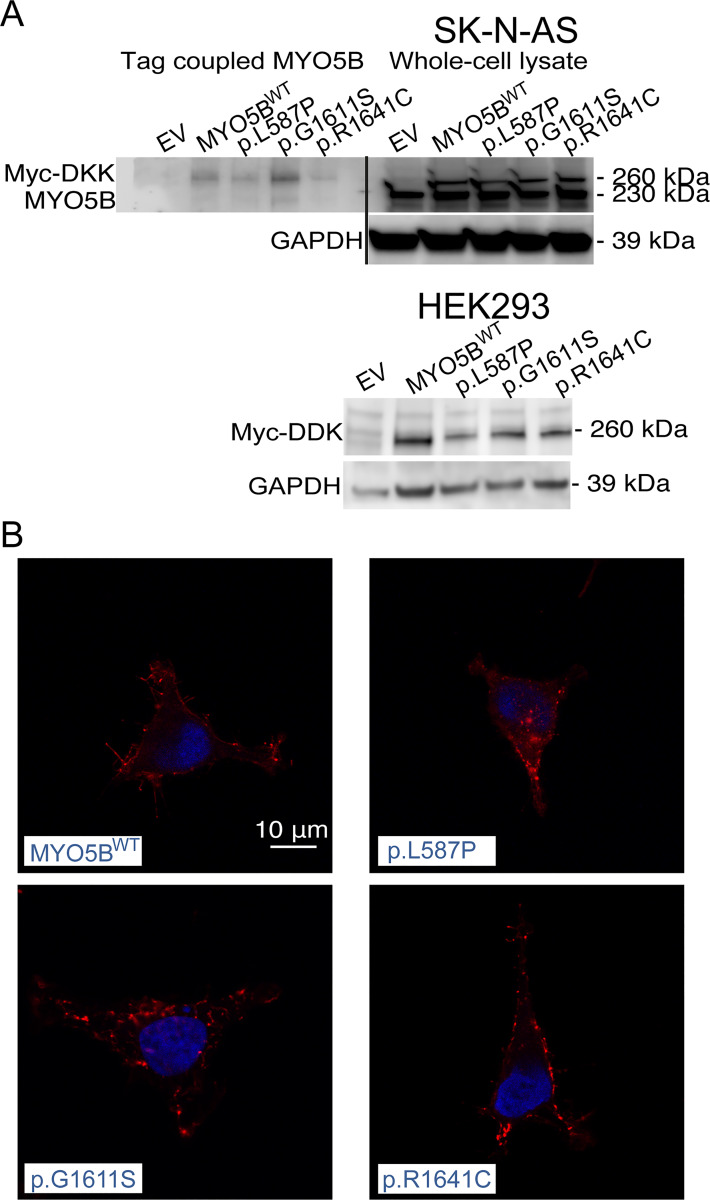
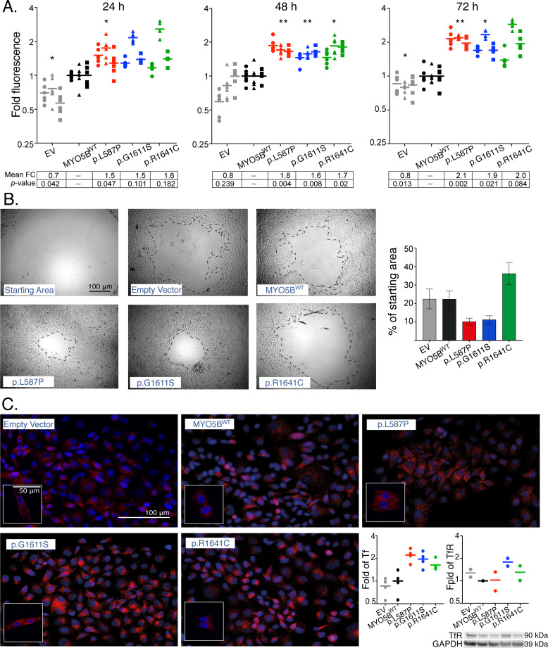
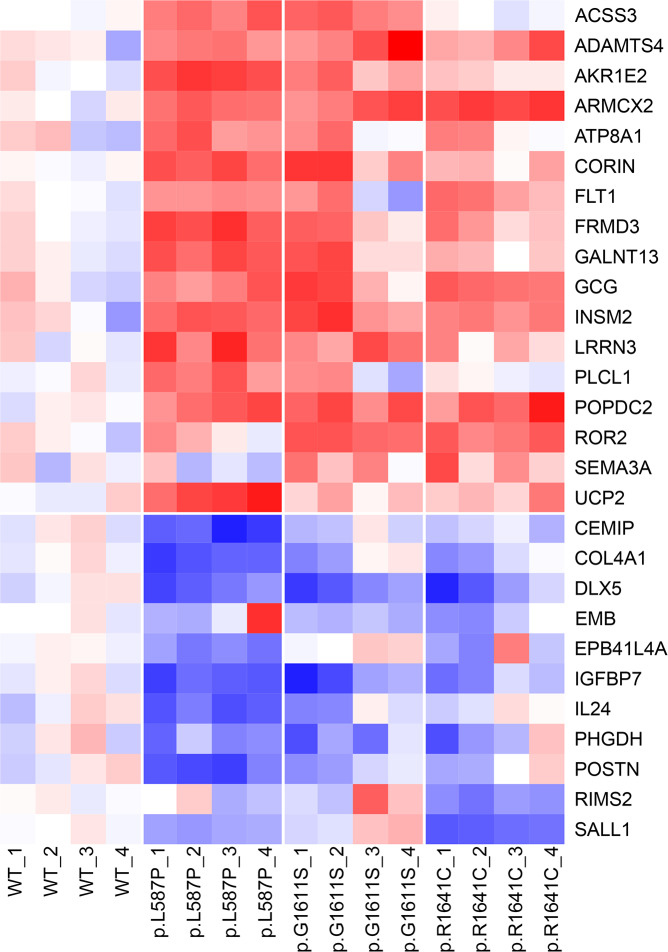
Identification of additional cancer-associated genes and secondary mutations driving the metastatic progression in pheochromocytoma and paraganglioma (PPGL) is important for subtyping, and may provide optimization of therapeutic regimens. We recently reported novel recurrent nonsynonymous mutations in the MYO5B gene in metastatic PPGL. Here, we explored the functional impact of these MYO5B mutations, and analyzed MYO5B expression in primary PPGL tumor cases in relation to mutation status. Immunohistochemistry and mRNA expression analysis in 30 PPGL tumors revealed an increased MYO5B expression in metastatic compared to non-metastatic cases. In addition, subcellular localization of MYO5B protein was altered from cytoplasmic to membranous in some metastatic tumors, and the strongest and most abnormal expression pattern was observed in a paraganglioma harboring a somatic MYO5B:p.G1611S mutation. In addition to five previously discovered MYO5B mutations, the present study of 30 PPGL (8 previous and 22 new samples) also revealed two, and hence recurrent, mutations in the gene paralog MYO5A. The three MYO5B missense mutations with the highest prediction scores (p.L587P, p.G1611S and p.R1641C) were selected and functionally validated using site directed mutagenesis and stable transfection into human neuroblastoma cells (SK-N-AS) and embryonic kidney cells (HEK293). In vitro analysis showed a significant increased proliferation rate in all three MYO5B mutated clones. The two somatically derived mutations, p.L587P and p.G1611S, were also found to increase the migration rate. Expression analysis of MYO5B mutants compared to wild type clones, demonstrated a significant enrichment of genes involved in migration, proliferation, cell adhesion, glucose metabolism, and cellular homeostasis. Our study validates the functional role of novel MYO5B mutations in proliferation and migration, and suggest the MYO5-pathway to be involved in the malignant progression in some PPGL tumors.


Conflict of interest statement

The authors have declared that no competing interests exist.

Figures

Fig 1
Fig 1. Localizations of mutations in MY05B.
Schematic presentation of the MYO5B gene, located on chromosome 18, containing 40 exons encoding the MYO5B protein (NP_001073936, 1848 amino acids). MYO5B constitutes of a myosin head domain and a globular tail (dilute) domain. Protein domains and motifs are presented according to Qiu et al [76], and the RAB8A- and RAB11A-binding sites according to Roland et al [23]. Localization of the five missense mutations in MYO5B identified in Wilzén et al [14] are marked in red, and the three selected mutations (p.L587P, p.G1611S, and p.R1641C) analyzed in the current study are marked with a star. Previously identified MYO5B mutations in colon cancer are according to Letellier et al [32] and marked in grey. IQ = IQ motif, BD = binding domain.
Fig 2
Fig 2. Expression of MYO5B in primary tumor tissue.
Immunohistochemistry (A) using MYO5B antibody of normal adrenal gland (top row), 3 representative non-metastatic PCC tumors (second row) and 6 representative PGL metastatic tumors (two bottom rows). Two tumors harboring somatic MYO5B mutations were included; CPN7 (p.L587P) and CPN8 (p.G1611S). Pictures are taken at 40 x magnification using a Nikon ECLIPSE E1000M microscope and a ProgRes C7 camera, scale bar shown is 100μm. mRNA expression analysis (B) by microarray in 26 PPGL cases. The normalized relative expression values are presented as fold change compared to the non-metastatic case with lowest expression (CPN2), and each tumor sample is presented with its mutation status and diagnosis; PCC (light grey), PGL (dark grey), non-metastatic (blue), metastatic (orange). The two cases harboring MYO5B mutations are marked with a star. NA = not available.
Fig 3
Fig 3. Expression and localization of MYO5B in cell lines.
Western blot in SK-N-AS cells (A) showing MYO5B expression in whole cell protein lysate and FLAG M2-antibody coupled lysate (n = 1, top panel). Western Blot using FLAG M2 antibody in HEK293 (n = 1, bottom panel). GAPDH used as loading control. Localization of Myc-DKK (B) in HEK293 (63X, LSM700) in pCMV6-MYO5B_L587P-Myc-DDK (p.L587P), pCMV6-MYO5B_G1611S-Myc-DDK (p.G1611S), and pCMV6-MYO5B_R1646C-Myc-DDK (p.R1641C). Myc-DKK is depicted in red and DAPI staining of nuclei in blue. Scale bar shown is 10μm.
Fig 4
Fig 4. Proliferation, migration and endosomal recycling in MYO5B-mutants.
Proliferation with CyQuant NF assay in SK-N-AS clones (A) shown as fold change at 24h, 48h, and 72h compared to mean MYO5B wild type (WT). Graphs show three independent experiments, each run in sextuplicates. Grey = empty vector, black = MYO5BWT, red = p.L587P, blue = p.G1611S, green = p.R1641C. Mean fold change (FC) and significance (p-value) compared to MYO5BWT within each time point are presented (lower panel). * p<0.05, ** p<0.01, paired t-test. Migration (Oris) in HEK293 clones (B) shown as representable photos from light microscope (5X) of cell area remaining in MYO5B-mutants (p.L587P, p.G1611S, and p.R1641C), MYO5BWT, and empty vector at 24 h compared to the starting wound area. Scale bar shown is 100μm. Graph show percent wound area left after 24 h (mean±SEM, n = 2) in each construct. Transferrin uptake assay in SK-N-AS clones (C), with transferrin stained in red and nuclei stained in blue (DAPI). Pictures are taken with 40X Zeiss Axioscope 2 Plus fluorescence microscope, and small inserts show representative pictures taken with 63X, LSM700 confocal microscope. Scale bars shown are 50 μm and 100μm, respectively. Graphs plot fold change of corrected total cell fluorescence (CTCF) of transferrin (Tf) uptake (from four fields of view taken with 40X), and Western Blot analysis of the Transferrin receptor (TfR) expression (from two independent experiments) in mutants and empty vector compared to MYO5BWT (n = 2). GAPDH was included as a loading control.
Fig 5
Fig 5. Heatmap of differentially expressed genes in MYO5B mutants.
The heat map is based on qPCR data from 28 genes presented as dCt-values normalized gene-wise to a standard deviation equal to one, and the mean of the MYO5BWT samples subtracted. Data include three MYO5B mutants (p.L587P, p.G1611S, p.R1641C) and MYO5B wild type (WT) run as four independent experiments (passages 1 = p21, 2 = p23, 3 = p28, 4 = p30 of SK-N-AS), analyzed at time point 48h in each proliferation experiment. Red = up-regulated, blue = down-regulated.

Similar articles

Cited by

References

    1. Wangberg B, Muth A, Khorram-Manesh A, Jansson S, Nilsson O, Forssell-Aronsson E, et al. Malignant pheochromocytoma in a population-based study: survival and clinical results. Ann N Y Acad Sci. 2006;1073:512–6. 10.1196/annals.1353.054 - DOI - PubMed
    1. Kolackov K, Tupikowski K, Bednarek-Tupikowska G. Genetic aspects of pheochromocytoma. Adv Clin Exp Med. 2012;21(6):821–9. - PubMed
    1. Goldstein RE, O'Neill JA Jr., Holcomb GW 3rd, Morgan WM 3rd, Neblett WW 3rd, Oates JA, et al. Clinical experience over 48 years with pheochromocytoma. Ann Surg. 1999;229(6):755–64; discussion 64–6. 10.1097/00000658-199906000-00001 - DOI - PMC - PubMed
    1. Fishbein L, Leshchiner I, Walter V, Danilova L, Robertson AG, Johnson AR, et al. Comprehensive Molecular Characterization of Pheochromocytoma and Paraganglioma. Cancer Cell. 2017;31(2):181–93. 10.1016/j.ccell.2017.01.001 - DOI - PMC - PubMed
    1. Hescot S, Leboulleux S, Amar L, Vezzosi D, Borget I, Bournaud-Salinas C, et al. One-year progression-free survival of therapy-naive patients with malignant pheochromocytoma and paraganglioma. J Clin Endocrinol Metab. 2013;98(10):4006–12. 10.1210/jc.2013-1907 - DOI - PubMed

Publication types