

More holes, more contrast? Comparing an 18-gauge non-fenestrated catheter with a 22-gauge fenestrated catheter for cardiac CT
- PMID: 32511272
- PMCID: PMC7279574
- DOI: 10.1371/journal.pone.0234311
More holes, more contrast? Comparing an 18-gauge non-fenestrated catheter with a 22-gauge fenestrated catheter for cardiac CT
Abstract
Objective: To compare the performance of an 18-gauge nonfenestrated catheter (18-NFC) with a 22-gauge fenestrated catheter (22-FC) for cardiac CT angiography (CCTA) in patients with suspected coronary heart disease.
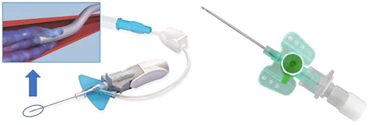
Subjects and methods: 74 consecutive patients imaged on a 2nd generation dual-source CT with arterial phase CCTA were included in this retrospective investigation to either an 18-NFC or 22-FC. In comparison to the 18-NFC, the 22-FC has three additional perforations for contrast agent dispersal proximal to the tip. We examined the two groups for differences in their average attenuation in the right and left ventricles (RV, LV) and in the atrium (RA, LA) as well as in the proximal right coronary artery (RCA) and the left main coronary artery (LM). The averages were calculated for both the 18-NFC and 22-FC.
Results: Catheters were successfully placed on the first attempt 97% (36/37) for 18-NFC and 95% (35/37) for the 22-FC. The following enhancement levels were measured: 22-FC (in Hounsfield-Units (HU)): RV = 203±29, LV = 523±36, RA = 198±29, LA = 519±38, RCA = 547±26, LM = 562±25; 18-NFC: RV = 146±26, LV = 464±32, RA = 141±24, LA = 438±35, RCA = 501±23, LM = 523±23; RV (p = 0,03), LV (p = 0.12), RA (p = 0.02), LA (p = 0.04), RCA (p = 0.3), LM (p = 0.33).
Conclusion: No significant differences in attenuation levels as well as in image quality of the coronary arteries were found between NFC and FC. Nevertheless, the 22-gauge FC examinations showed significantly higher attenuation in the left and right atrium as well as the right ventricle. Patients with poor venous access may benefit from a smaller gauge catheter that can deliver sufficiently high flow rates for CCTA.
Conflict of interest statement
Dr. Schoenberg reports that the institute of clinical radiology and nuclear medicine has research agreements with Siemens Healthcare GmbH. Dr. Schoepf receives institutional research support from Astellas, Bayer, and Siemens. Dr. Schoepf has received consulting fees and or speaker honoraria from Bayer, Elucid BioImaging, GE, Guerbet, HeartFlow Inc., and Siemens. UMCG receives institutional research support from Siemens. The other authors have no conflict of interest to disclose. This does not alter our adherence to PLOS ONE policies on sharing data and materials.
Figures


References
-
- Silvennoinen HM, Hamberg LM, Lindsberg PJ, Valanne L, Hunter GJ. CT perfusion identifies increased salvage of tissue in patients receiving intravenous recombinant tissue plasminogen activator within 3 hours of stroke onset. AJNR Am J Neuroradiol. 2008;29(6):1118–23. Epub 2008/04/12. 10.3174/ajnr.A1039 . - DOI - PMC - PubMed
-
- Goshima S, Kanematsu M, Nishibori H, Sakurai K, Miyazawa D, Watanabe H, et al. CT of the pancreas: comparison of anatomic structure depiction, image quality, and radiation exposure between 320-detector volumetric images and 64-detector helical images. Radiology. 2011;260(1):139–47. Epub 2011/03/17. 10.1148/radiol.11101459 . - DOI - PubMed
Publication types
MeSH terms
Substances
LinkOut - more resources
Full Text Sources
