. 2020 Jun 8;16(6):e1008587.
doi: 10.1371/journal.ppat.1008587. eCollection 2020 Jun.

Three-dimensional ultrastructure of Plasmodium falciparum throughout cytokinesis

Affiliations

Three-dimensional ultrastructure of Plasmodium falciparum throughout cytokinesis

Rachel M Rudlaff et al. PLoS Pathog. .

Abstract

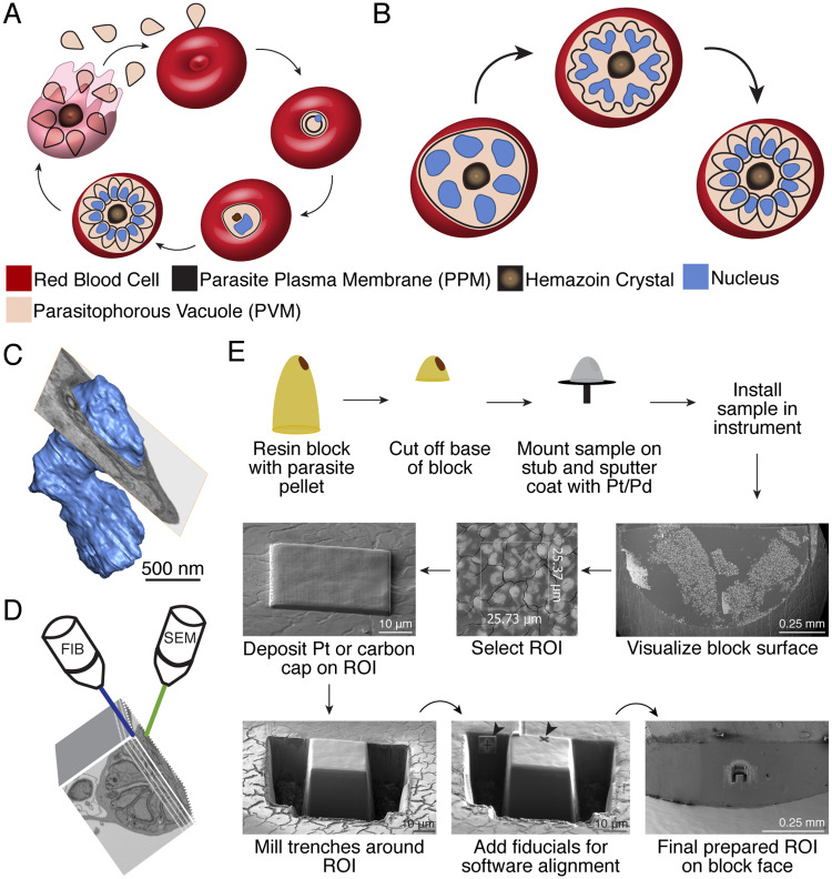
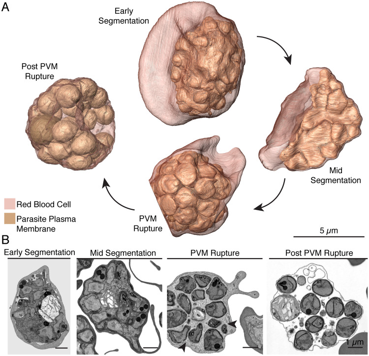
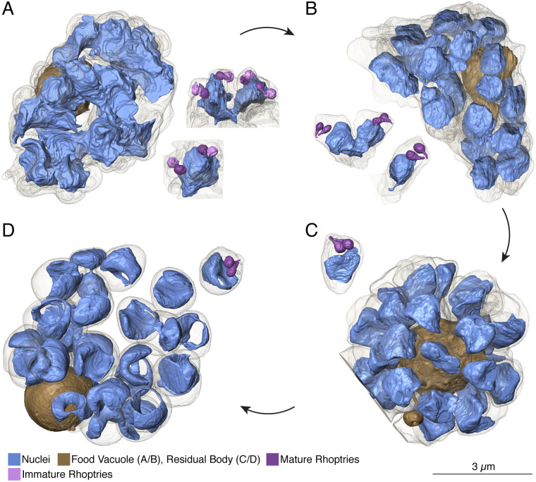
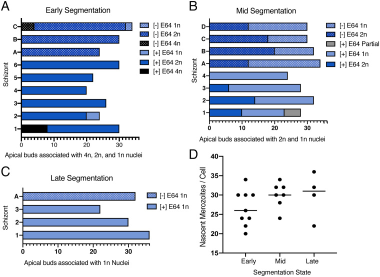
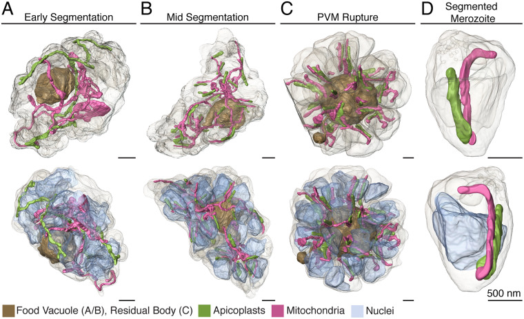
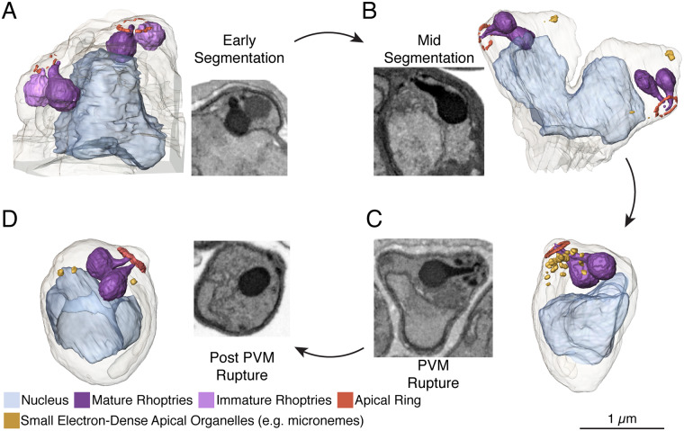
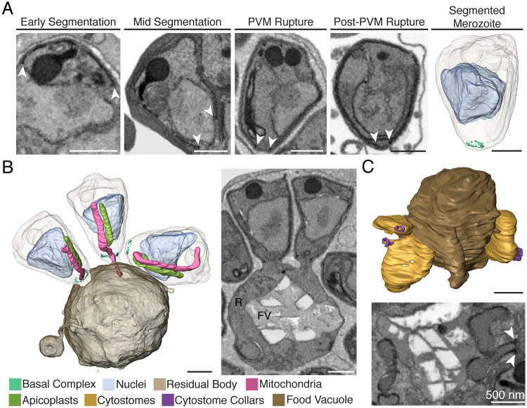
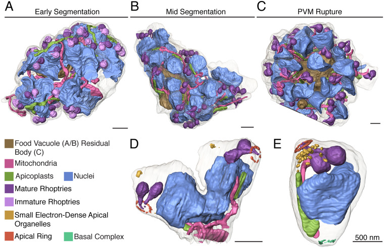
New techniques for obtaining electron microscopy data through the cell volume are being increasingly utilized to answer cell biologic questions. Here, we present a three-dimensional atlas of Plasmodium falciparum ultrastructure throughout parasite cell division. Multiple wild type schizonts at different stages of segmentation, or budding, were imaged and rendered, and the 3D structure of their organelles and daughter cells are shown. Our high-resolution volume electron microscopy both confirms previously described features in 3D and adds new layers to our understanding of Plasmodium nuclear division. Interestingly, we demonstrate asynchrony of the final nuclear division, a process that had previously been reported as synchronous. Use of volume electron microscopy techniques for biological imaging is gaining prominence, and there is much we can learn from applying them to answer questions about Plasmodium cell biology. We provide this resource to encourage readers to consider adding these techniques to their cell biology toolbox.


Conflict of interest statement

The authors have declared that no competing interests exist.

Figures

Fig 1
Fig 1. Plasmodium falciparum asexual life cycle and FIB-SEM sample preparation.
A. A merozoite invades a red blood cell (RBC) to initiate infection (top center). Post-invasion, the parasite develops into a biconcave early trophozoite known as a ring inside its parasitophorous vacuole (PV) (right). The parasite becomes more metabolically active and begins to replicate its DNA as a late trophozoite (bottom right). As a schizont (bottom left) the parasite produces nuclei and organelles for its daughter cells, then undergoes cytokinesis to package these contents into individual merozoites. Egress (left) releases merozoites from the PV and RBC to invade a new RBC. B. In schizogony, the parasite produces DNA and organelles for its daughter cells asynchronously within a common cytoplasm (left). During segmentation, the parasite plasma membrane invaginates around nascent daughter cell buds (middle) to form fully segmented merozoites (right). This model depicts the hypothesis that the final round of karyokinesis is synchronous and simultaneous with cytokinesis. C. Volume electron microscopy reveals features not captured in single slices. A slice of EM data shown as an ortho-slice through the rendered nucleus, which contains subsurface features unseen in the single slice shown. Scale bar interpolated from structure sizes in EM data D. FIB-SEM employs two beams to serially mill the face off a resin block with a focused ion beam, then image with a scanning electron beam. In some instruments, milling and imaging can be performed simultaneously. E. FIB-SEM workflow: A resin block (yellow) with embedded parasite pellet (brown) is shaved down to reduce block height, mounted on a SEM stub with carbon tape, and sputter coated with platinum-palladium (Pt/Pd). The sample is installed in the FIB-SEM instrument and block surface visualized to identify regions of cell density. A 1–2 μm platinum (shown) or carbon (for data captured in this resource) cap is applied to the region of interest (ROI) to improve conductivity. Trenches are milled to expose the front of the block face for imaging and fiducials (arrows) are milled for focusing by the Atlas control software. For the data presented in this resource, fiducials were instead sandwiched between layers of the carbon cap.
Fig 2
Fig 2. Parasite plasma membrane dynamics throughout segmentation.
A. Renderings of the four different schizont stages inside their host red blood cells through segmentation. Scale bars of renderings interpolated from structure sizes in EM data. At early segmentation, the mother parasite plasma membrane begins to invaginate, forming buds for each nascent merozoite that slightly protrude from the mother parasite mass. At mid-segmentation, these buds are more pronounced and jut out of the mother parasite. By PVM-rupture, the parasite plasma membrane cloaks each new merozoite, which is shaped like an inverted cone. Post-PVM rupture, merozoites fill the red blood cell and take on a more spherical shape. B. Single SEM images of the four schizonts chosen for rendering. We note that for the early segmentation schizont, the increased contrast between the parasite plasma membrane and parasitophorous vacuole is an artifact of sample preparation and provides additional contrast helpful for rendering. Arrows indicate region of PVM rupture.
Fig 3
Fig 3. Nuclear architecture throughout segmentation.
A. In early segmentation, nuclei appear jagged and are associated with two (in 11 of 13) or four (in 2 of 13) sets of rhoptries. The mature rhoptry from each pair is in close association with the nucleus. B. In mid-segmentation, nuclei have a smoother appearance, and of fully rendered nuclei, five are 2n and 13 are 1n. Half of imaged rhoptry sets retain a nuclear association. C. By PVM rupture, all nuclei are separated into their respective merozoites. No rhoptry sets have a visible nuclear association. D. Post-PVM rupture, nuclei have depressed centers, forming a toroid shape or C-shape. We note that this may be due to prolonged RBC entrapment with E64. One daughter cell was ruptured in sample preparation, resulting in two lobes of bridged DNA (seen near the top of the cell). In A and B, the food vacuole is rendered, in C and D, the residual body, containing the food vacuole and residual cytoplasmic material, is rendered. Scale bars of renderings interpolated from structure sizes in EM data.
Fig 4
Fig 4. Nuclear status of parasites at early, mid, and late segmentation.
A. Graph demonstrating the number of apical buds associated with 4n, 2n, and 1n nuclei in three schizonts from an E64-untreated block and six schizonts from an E64 treated block. B. Graph demonstrating the number of apical buds associated with 2n and 1n nuclei in four schizonts from a [–]E64 (A-D) and four schizonts from a [+]E64 (1–4) treated sample. C. Graph depicting the number of apical buds associated with 1n nuclei in one [–]E64 treated schizont and three [+]E64 treated schizonts. D. Plot of the number of nascent merozoites per fully captured cell. Line placed at median for each set.
Fig 5
Fig 5. Apicoplast and mitochondria morphology throughout segmentation.
A. In early segmentation, the apicoplast and mitochondrion each form a sinuous structure throughout the parasite. B. By mid-segmentation, apicoplasts have divided to form one organelle for each nascent daughter cell and are closely associated with the mitochondrion, which remains undivided. C. At PVM rupture, both apicoplasts and mitochondria are divided and are largely situated within segmented daughter cells. D. A segmented merozoite from the schizont in (C), with a single mitochondrion and apicoplast. Scale bars of renderings interpolated from structure sizes in EM data.
Fig 6
Fig 6. Apical end development throughout schizogony.
A. Apical end of a 2n nucleus in early segmentation. At this stage, the apical ring is not well-defined, resulting in fragmented rendering. Each pair of rhoptries has a mature, electron-dense bulb, and a less mature, less electron-dense bulb, visible in the electron micrograph inset and rendered in darker and lighter shades of purple, respectively. B. At mid-segmentation, the apical ring is more defined and both rhoptries in each set are mature. A few small, electron-dense apical organelles begin to appear. C. At PVM rupture, the apical head of the parasite is mature, with a well-defined apical ring, multiple small electron-dense organelles, and mature rhoptries. D. Post-PVM rupture, fewer apical organelles remain, presumably due to discharge of a subset of microneme-related organelles, but the defined apical ring and rhoptries are not materially different from the PVM rupture schizont. The three apical polar rings are visible in electron micrographs for each daughter cell, but the first two are too thin to be rendered. Electron micrograph insets for A-C do not correspond to the rendered apical head. Scale bars of renderings interpolated from structure sizes in EM data.
Fig 7
Fig 7. The basal end and food vacuole.
A. The basal ring resides at the teardrop shaped basal end of the IMC (white arrows). At PVM rupture, this structure is defined enough to render. B. At PVM rupture, eight merozoites remained attached to the residual body. Two attached and one unattached merozoites are rendered, including one merozoite with its mitochondrion connected to the residual body. R = residual body, FV = food vacuole. C. At mid-segmentation, we visualize the parasite taking up RBC cytosol at cytostomes. These invaginations are located near the food vacuole and their necks are encircled in cytostomal collars, delineated by an electron-dense ring (white arrows). Scale bars of renderings interpolated from structure sizes in EM data.
Fig 8
Fig 8. The whole schizont.
A. In early segmentation, sets of rhoptries composed of one mature and one immature bulb are associated with 4n or 2n nuclei. The apicoplast and mitochondrion twist through the cell, undivided. B. At mid-segmentation, pairs of rhoptries are fully mature and nuclei are either 2n or 1n. Apicoplasts are divided and associated with the undivided mitochondrion. C. At PVM rupture, individual sets of rhoptries, nuclei, apicoplasts, and mitochondria are separated into their respective merozoite plasma membranes. Most merozoites are fully separated, but eight remain attached to the residual body. D. A mid-segmentation bud with material for two daughter cells. The mitochondrion snakes out of the rendered bud to connect with other budding cells. E. A fully segmented merozoite with a mature apical head, a segmented nucleus, apicoplast, mitochondrion, and the basal ring. Scale bars of renderings interpolated from structure sizes in EM data.

Similar articles

Cited by

References

    1. Farrar J, Hotez PJ, Junghanss T, Kang G, Lalloo D, White NJ. Manson’s Tropical Diseases E-Book: Elsevier Health Sciences; 2013.
    1. Cowman AF, Healer J, Marapana D, Marsh K. Malaria: Biology and Disease. Cell. 2016;167(3):610–24. Epub 2016/10/22. 10.1016/j.cell.2016.07.055 . - DOI - PubMed
    1. van Dooren GG, Marti M, Tonkin CJ, Stimmler LM, Cowman AF, McFadden GI. Development of the endoplasmic reticulum, mitochondrion and apicoplast during the asexual life cycle of Plasmodium falciparum. Mol Microbiol. 2005;57(2):405–19. Epub 2005/06/28. 10.1111/j.1365-2958.2005.04699.x . - DOI - PubMed
    1. Read M, Sherwin T, Holloway SP, Gull K, Hyde JE. Microtubular organization visualized by immunofluorescence microscopy during erythrocytic schizogony in Plasmodium falciparum and investigation of post-translational modifications of parasite tubulin. Parasitology. 1993;106 (Pt 3):223–32. Epub 1993/04/01. 10.1017/s0031182000075041 . - DOI - PubMed
    1. Francia ME, Striepen B. Cell division in apicomplexan parasites. Nat Rev Microbiol. 2014;12(2):125–36. Epub 2014/01/05. 10.1038/nrmicro3184 . - DOI - PubMed

Publication types

MeSH terms