[Preprint]. 2020 Apr 21.
doi: 10.26434/chemrxiv.12153615.

Structural Similarity of SARS-CoV2 Mpro and HCV NS3/4A Proteases Suggests New Approaches for Identifying Existing Drugs Useful as COVID-19 Therapeutics

Affiliations

Structural Similarity of SARS-CoV2 Mpro and HCV NS3/4A Proteases Suggests New Approaches for Identifying Existing Drugs Useful as COVID-19 Therapeutics

Khushboo Bafna et al. ChemRxiv. .

Update in

Abstract

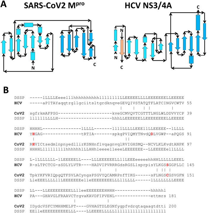
During the current COVID-19 pandemic more than 160,000 people have died worldwide as of mid-April 2020, and the global economy has been crippled. Effective control of the SARS-CoV2 virus that causes the COVID-19 pandemic requires both vaccines and antivirals. Antivirals are particularly crucial to treat infected people during the period of time that an effective vaccine is being developed and deployed. Because the development of specific antiviral drugs can take a considerable length of time, an important approach is to identify existing drugs already approved for use in humans which could be repurposed as COVID-19 therapeutics. Here we focus on antivirals directed against the SARS-CoV2 Mpro protease, which is required for virus replication. A structural similarity search showed that the Hepatitis C virus (HCV) NS3/4A protease has a striking three-dimensional structural similarity to the SARS-CoV2 Mpro protease, particularly in the arrangement of key active site residues. We used virtual docking predictions to assess the hypothesis that existing drugs already approved for human use or clinical testing that are directed at the HCV NS3/4A protease might fit well into the active-site cleft of the SARS-CoV2 protease (Mpro). AutoDock docking scores for 12 HCV protease inhibitors and 9 HIV-1 protease inhibitors were determined and compared to the docking scores for an α-ketoamide inhibitor of Mpro, which has recently been shown to inhibit SARS-CoV2 virus replication in cell culture. We identified eight HCV protease inhibitors that bound to the Mpro active site with higher docking scores than the α-ketoamide inhibitor, suggesting that these protease inhibitors may effectively bind to the Mpro active site. These results provide the rationale for us to test the identified HCV protease inhibitors as inhibitors of the SARS-CoV2 protease, and as inhibitors of SARS-CoV2 virus replication. Subsequently these repurposed drugs could be evaluated as COVID-19 therapeutics.

Keywords: COVID-19; Drug Discovery; Hepatitis C Virus (HCV) NS3/4A protease inhibitors; Human Immunodeficiency Virus 1 (HIV-1) protease inhibitors; Severe Acute Respiratory Syndrome (SARS); structural bioinformatics.


Conflict of interest statement

Conflict of Interest Statement: G.T.M. is a founder of Nexomics Biosciences, Inc.

Figures

Fig. 1.
Fig. 1.. X-ray crystal structures of viral proteases.
A) SARS-CoV2 Mpro (PDB id 6Y2G), B) HCV protease NS3/4A (PDB id 2P59), and C) HIV-1 protease (PDB id 4LL3). HCV NS3/4A protease and SARS-CoV2 Mpro both have a double β-barrel fold architecture, with a substrate binding site located in a shallow cleft between two antiparallel β-barrels (shown in cyan and blue). Only one protomer is shown for A and B. The α-helical domain III of Mpro is shown in green. The HIV protease has a different fold architecture, and the substrate binding site is located in between the two protomers. For the HIV-1 protease both protomers are shown. The structures of bound inhibitors in these crystal structures are illustrated as magenta sticks.
Fig. 2.
Fig. 2.. Superimposition of viral proteases.
The backbone structure of the SARS-CoV2 Mpro, PDB 6Y2G (green) is superimposed on the backbone structure of hepatitis C virus protease NS3/4A, PDB 2P59 (cyan). The regions identified by DALI (18) as structurally-analogous are shown in color (green and cyan), and the regions that are not structurally-analogous are shown in gray. This superimposition of backbone atoms results in superimposition of the catalytic residues Cys145 and His41 of the SARS-CoV2 Mpro with Ser139 and His57 of HCV protease. Residue Asp81 of the HCV protease catalytic triad is also shown.
Fig. 3.
Fig. 3.. Comparison of fold topology diagrams and structure-based sequence alignments of SARS-CoV2 Mpro and HCV NS3/4A proteases.
A) Fold topology diagrams demonstrate similar fold architectures but different topologies. B) The DALI server (18) provides a structure-based sequence alignment of HCV NS3/4A (HCV) and SARS-CoV2 Mpro (CoV2). Catalytic residues of HCV NS3/4A (His57, Asp81 and Ser139) and SARS-Cov2 Mpro (His41 and Cys145) are highlighted in bold red. Three-state secondary structure definitions by DSSP (19) (H=helix, E=sheet, L=coil) are shown above each amino acid sequence. Structurally equivalent residues are in uppercase, structurally non-equivalent residues (e.g. in loops) are in lowercase. Amino acid identities are marked by vertical bars. The structure-based alignment results in alignment of key catalytic residues His41 and Cys145 of the SARS-CoV2 Mpro with His57 and Ser139 of HCV NS3/4A protease.
Fig. 4.
Fig. 4.. Docking of α-ketoamide protease inhibitor 13b (13) to SARS-CoV2 Mpro.
A) Grid box placed around reference ligand 13b to define the binding site. B) Lowest energy AutoDock pose using a rigid conformation of α-ketoamide inhibitor 13b, compared with the bound-state ligand conformation observed in the X-ray crystal structure of the complex (score = −7.19 kcal/mol). C) Lowest energy AutoDock pose observed among 100 docking simulations (score = −9.17 kcal/mol). D) Lowest energy AutoDock pose that is most similar to the conformation seen in the crystal structure (score = −9.03 kcal/mol). In each of panels B-D, the Mpro protein is shown in surface representation (gray), together with the X-ray crystal structure of α-ketoamide inhibitor 13b bound in the active site of Mpro (green sticks), and the predicted AutoDock conformation (magenta sticks).
Fig. 5.
Fig. 5.. Docking of HCV protease NS3/4A inhibitor drugs to SARS CoV2 Mpro .
Top panels - Molecular structures of two HCV protease inhibitor drugs. Middle panels – Lowest energy AutoDock pose of these HCV protease inhibitors (magenta sticks) in the SARS CoV2 Mpro active site, compared to the pose observed in the crystal structure (PDB id 6Y2G) available for the SARS-CoV2 Mpro α-ketoamide inhibitor 13b (green sticks). Bottom panels – Details of atomic interactions in the lowest energy AutoDock poses of these HCV protease inhibitors. Hydrogen bonds and hydrophobic interactions between the drug and the enzyme are shown with yellow solid lines and black dashed lines, respectively. Sidechains of catalytic residues His41 and Cys145 are labeled, along with other protein residues that form key interactions with these drugs.
Fig. 6.
Fig. 6.. Docking of HCV protease NS3/4A inhibitor drugs to SARS CoV2 Mpro .
Top panels - Molecular structures of two HCV protease inhibitor drugs. Middle panels – Lowest energy AutoDock pose of these HCV protease inhibitors (magenta sticks) in the SARS CoV2 Mpro active site, compared to the pose observed in the crystal structure (PDB id 6Y2G) available for the SARS-CoV2 Mpro α-ketoamide inhibitor 13b (green sticks). Bottom panels – Details of atomic interactions in the lowest energy AutoDock poses of these HCV protease inhibitors. Hydrogen bonds and hydrophobic interactions between the drug and the enzyme are shown with yellow solid lines and black dashed lines, respectively. Sidechains of catalytic residues His41 and Cys145 are labeled, along with other protein residues that form key interactions with these drugs.
Fig. 7.
Fig. 7.. Docking of HCV protease NS3/4A inhibitor drugs to SARS CoV2 Mpro .
Top panels - Molecular structures of two HCV protease inhibitor drugs. Middle panels – Lowest energy AutoDock pose of these HCV protease inhibitors (magenta sticks) in the SARS CoV2 Mpro active site, compared to the pose observed in the crystal structure (PDB id 6Y2G) available for the SARS-CoV2 Mpro α-ketoamide inhibitor 13b (green sticks). Bottom panels – Details of atomic interactions in the lowest energy AutoDock poses of these HCV protease inhibitors. Hydrogen bonds and hydrophobic interactions between the drug and the enzyme are shown with yellow solid lines and black dashed lines, respectively. Sidechains of catalytic residues His41 and Cys145 are labeled, along with other protein residues that form key interactions with these drugs.
Fig. 8.
Fig. 8.. Docking of HCV protease NS3/4A inhibitor drugs to SARS CoV2 Mpro .
Top panels - Molecular structures of two HCV protease inhibitor drugs. Middle panels – Lowest energy AutoDock pose of these HCV protease inhibitors (magenta sticks) in the SARS CoV2 Mpro active site, compared to the pose observed in the crystal structure (PDB id 6Y2G) available for the SARS-CoV2 Mpro α-ketoamide inhibitor 13b (green sticks). Bottom panels – Details of atomic interactions in the lowest energy AutoDock poses of these HCV protease inhibitors. Hydrogen bonds and hydrophobic interactions between the drug and the enzyme are shown with yellow solid lines and black dashed lines, respectively. Sidechains of catalytic residues His41 and Cys145 are labeled, along with other protein residues that form key interactions with these drugs.

References

    1. De Wit E., Van Doremalen N., Falzarano D. and Munster V. J. SARS and MERS: Recent insights into emerging coronaviruses. Nat Rev Microbiol 2016, 14: 523–534. 10.1038/nrmicro.2016.81 - DOI - PMC - PubMed
    1. Zhou P., Yang X. L., Wang X. G., Hu B., Zhang L., Zhang W., Si H. R., Zhu Y., Li B., Huang C. L., et al. A pneumonia outbreak associated with a new coronavirus of probable bat origin. Nature 2020, 579: 270–273. 10.1038/s41586-020-2012-7 - DOI - PMC - PubMed
    1. Wu F., Zhao S., Yu B., Chen Y. M., Wang W., Song Z. G., Hu Y., Tao Z. W., Tian J. H., Pei Y. Y., et al. A new coronavirus associated with human respiratory disease in China. Nature 2020, 579: 265–269. 10.1038/s41586-020-2008-3 - DOI - PMC - PubMed
    1. Gordon D. E., Jang G. M., Bouhaddou M., Xu J., Obernier K., O’meara M. J., Guo J. Z., Swaney D. L., Tummino T. A., Huettenhain R., et al. A SARS-CoV-2-human protein-protein interaction map reveals drug targets and potential drug-repurposing. bioRxiv 2020. 10.1101/2020.03.22.002386 - DOI - PMC - PubMed
    1. Liu X. and Wang X. J. Potential inhibitors against 2019-nCoV coronavirus M protease from clinically approved medicines. J Genet Genomics 2020, 47: 119–121. 10.1016/j.jgg.2020.02.001 - DOI - PMC - PubMed

Publication types

LinkOut - more resources