[Preprint]. 2020 Oct 6:2020.04.17.047480.
doi: 10.1101/2020.04.17.047480.

Coronavirus infection and PARP expression dysregulate the NAD Metabolome: an actionable component of innate immunity

Affiliations

Coronavirus infection and PARP expression dysregulate the NAD Metabolome: an actionable component of innate immunity

Collin D Heer et al. bioRxiv. .

Update in

Abstract

Poly-ADP-ribose polymerase (PARP) superfamily members covalently link either a single ADP-ribose (ADPR) or a chain of ADPR units to proteins using nicotinamide adenine dinucleotide (NAD) as the source of ADPR. While the well-known poly-ADP-ribosylating (PARylating) PARPs primarily function in the DNA damage response, many non-canonical mono-ADP-ribosylating (MARylating) PARPs are associated with cellular antiviral responses. We recently demonstrated robust upregulation of several PARPs following infection with Murine Hepatitis Virus (MHV), a model coronavirus. Here we show that SARS-CoV-2 infection strikingly upregulates MARylating PARPs and induces the expression of genes encoding enzymes for salvage NAD synthesis from nicotinamide (NAM) and nicotinamide riboside (NR), while downregulating other NAD biosynthetic pathways. We show that overexpression of PARP10 is sufficient to depress cellular NAD and that the activities of the transcriptionally induced enzymes PARP7, PARP10, PARP12 and PARP14 are limited by cellular NAD and can be enhanced by pharmacological activation of NAD synthesis. We further demonstrate that infection with MHV induces a severe attack on host cell NAD+ and NADP+. Finally, we show that NAMPT activation, NAM and NR dramatically decrease the replication of an MHV virus that is sensitive to PARP activity. These data suggest that the antiviral activities of noncanonical PARP isozyme activities are limited by the availability of NAD, and that nutritional and pharmacological interventions to enhance NAD levels may boost innate immunity to coronaviruses.

Keywords: ADP-ribosylation; COVID-19; Severe acute respiratory syndrome coronavirus 2 (SARS-CoV-2); interferon; nicotinamide adenine dinucleotide (NAD); poly(ADP-ribose) polymerase (PARP); transcriptomics.


Conflict of interest statement

COMPETING INTERESTS CB is chief scientific adviser of ChromaDex and owns shares of ChromaDex stock. CB, SAJT, SP and ARF filed an invention disclosure on uses of NAD-boosting with respect to protection against coronavirus infection. Others declare no competing interests.

Figures
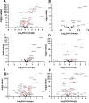
Figure 1.
Figure 1.. SARS-CoV-2 dysregulates the NAD gene set in vitro as a function of viral load.
Differential expression analysis was performed on RNAseq data with respect to a 71 gene set representing the NAD transcriptome (Supplementary Table 1). Depicted are Volcano Plots representing normalized relative expression versus -log(P) with respect to mock infected in A) human Calu3 lung cancer cells (MOI = 2), B) NHBE cells (MOI = 2), C and D), A549 cells at low MOI without and with introduction of ACE2 expression, respectively, E and F) A549 cells at high MOI = 2 without and with introduction of ACE2 expression. Further information is available in Supplementary Materials 2–7.
Figure 2.
Figure 2.. SARS-CoV-2 dysregulates the NAD gene set in vivo.
Differential expression analysis was performed on RNAseq data with respect to mock infected in A) expanding enterocytes (MOI=1), B) ferret trachea infected with SARS-CoV-2, C) lung of a diseased COVID-19 patient versus a control lung sample and D) BALF from SARS-CoV2 infected versus healthy control human patients. Further information is available in Supplementary Materials 8–11.
Figure 3.
Figure 3.. PARP10 overexpression is sufficient to depress cellular NAD+ levels while SBI enhances activities of overexpressed PARP7, PARP10, PARP12 and PARP14.
A) HEK 293T cells were grown with the indicated expression plasmids for GFP or PARP10 and treated with NAMPT activator (10 μM SBI). n = 3 for each group. Error bars represent SEM, p-values are from an unpaired two-tailed t-test. See also Supplementary Information 12. B) GFP, PARP7, PARP10, PARP12 and PARP14- expressing HEK293 cells were treated with 10 μM SBI and cells were collected 18 hours later. Western blot using indicated antibodies indicate that SBI promotes PARP7, PARP10, PARP12 and PARP14 activity. n=3. Representative blots of three independent experiments are shown. ** p ≤ 0.01, *** p ≤ 0.001
Figure 4.
Figure 4.. MHV infection disturbs the NAD metabolome.
A) DBT cells and B) BMDM cells were mock infected or infected with MHV at a MOI of 3 PFU/cell and cells were collected at 12 hrs post-infection. n = 3–4 Mock; n = 4 MHV. Error bars represent SEM, p-values are from unpaired two-tailed t-test. **p ≤ 0.01, ***p ≤ 0 .001. See also Supplementary Materials 13–14.
Figure 5.
Figure 5.. Boosting NAD+ levels depresses replication of CARH mutant MHV.
A) NAD biosynthetic pathways. Red arrows depict gene expression that is depressed by SARS-CoV-2. Green arrows depict gene expression that is increased by SARS-CoVo2. B) 17Cl-1 cells were infected with 0.1 PFU/cell WT or N1347A MHV and either mock treated (DMSO or H2O) or treated with NA, NAM, SBI or NR as described in Methods. DMSO served as a solvent control for NAM, SBI and NA, while H2O served as a solvent control for NR. Cells were collected at 18 hpi and analyzed for virus replication by plaque assay. Data are representative of two independent experiments, n = 3 biological replicates. C) BMDMs were infected with 0.1 PFU/cell WT or N1347A MHV and treated with H2O or treated with NR as described in Methods. Cells were collected at 18 hpi and analyzed for virus replication by plaque assay. Data are representative of two independent experiments. n = 4 biological replicates. *p ≤ 0.05, **p ≤ 0.01, ***p ≤ 0.001.

References

    1. Dong E., Du H., and Gardner L. (2020) An interactive web-based dashboard to track COVID-19 in real time. Lancet Infect Dis 20, 533–534 - PMC - PubMed
    1. Wu D., Wu T., Liu Q., and Yang Z. (2020) The SARS-CoV-2 outbreak: what we know. International journal of infectious diseases : IJID : official publication of the International Society for Infectious Diseases 94, 44–48 - PMC - PubMed
    1. Fehr A. R., and Perlman S. (2015) Coronaviruses: an overview of their replication and pathogenesis. Methods Mol Biol 1282, 1–23 - PMC - PubMed
    1. Zhu N., Zhang D., Wang W., Li X., Yang B., Song J., Zhao X., Huang B., Shi W., Lu R., Niu P., Zhan F., Ma X., Wang D., Xu W., Wu G., Gao G. F., Tan W., China Novel Coronavirus, I., and Research, T. (2020) A Novel Coronavirus from Patients with Pneumonia in China, 2019. N Engl J Med 382, 727–733 - PMC - PubMed
    1. Wu C., Liu Y., Yang Y., Zhang P., Zhong W., Wang Y., Wang Q., Xu Y., Li M., Li X., Zheng M., Chen L., and Li H. (2020) Analysis of therapeutic targets for SARS-CoV-2 and discovery of potential drugs by computational methods. Acta Pharm Sinica B 10, 766–788 - PMC - PubMed

Publication types