

This is a preprint.
It has not yet been peer reviewed by a journal.
The National Library of Medicine is
running a pilot
to include preprints that result from research funded by NIH in PMC and PubMed.
[Preprint]. 2020 Mar 31:2020.03.29.008631.
doi: 10.1101/2020.03.29.008631.
Azithromycin and ciprofloxacin have a chloroquine-like effect on respiratory epithelial cells
Affiliations
- PMID: 32511331
- PMCID: PMC7239066
- DOI: 10.1101/2020.03.29.008631
Item in Clipboard
Azithromycin and ciprofloxacin have a chloroquine-like effect on respiratory epithelial cells
bioRxiv.
.
Abstract
There is interest in the use of chloroquine/hydroxychloroquine (CQ/HCQ) and azithromycin (AZT) in COVID-19 therapy. Employing cystic fibrosis respiratory epithelial cells, here we show that drugs AZT and ciprofloxacin (CPX) act as acidotropic lipophilic weak bases and confer in vitro effects on intracellular organelles similar to the effects of CQ. These seemingly disparate FDA-approved antimicrobials display a common property of modulating pH of endosomes and trans-Golgi network. We believe this may in part help understand the potentially beneficial effects of CQ/HCQ and AZT in COVID-19, and that the present considerations of HCQ and AZT for clinical trials should be extended to CPX.
Figures

A. AZT chemical formula and pK of its amino groups. B-G. Cystic fibrosis (IB3–1 or primary bronchial epithelial cells from CF lung transplants) and normal (S9, CFTR-corrected IB3–1 cells) were transfected with either TGN38-GFP pHluorin (B-D) or cellubrevin-GFP pHluorin (E-G) and pH of TGN (B-D) or recycling endosome (E-G) determined ratiometrically (emission intensity at 508 nm upon excitation at 410 nm and 470 nm) with or without AZT treatment. Images in B and E: top row, untreated primary bronchial CF cell; bottom row, primary bronchial CF cell treated with AZT. Color look-up table, R (410/470) ratio of emission intensity at 508 nm upon illumination at 410 and 470 nm. C, TGN pH in IB3–1 (CF) cells with or without treatment with AZT, or in S9 (CFTR-corrected IB3–1 cells). D, correction of TGN pH in primary CF respiratory epithelial cells by AZT. E, ratiometric fluorescence images with cellubrevin-GFP pHluorin in primary CF respiratory epithelial cells from lung transplant. F, correction with AZT of cellubrein endosome pH in CF cells (IB3–1). G. Correction with AZT of pH in the cellubrevin endosomes in primary CF respiratory epithelial cells, lung transplant, P (ANOVA), * <0.05; ** <0.01, n=6.
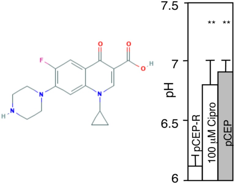
Phenotypically CF cells (pCEP-R) were transfected with TGN38-GFP pHlourin. Transfected cells were treated with 10 μM CPX for 1 h and pH determined ratiometrically as in studies with AZT. pCEP, normal cells.
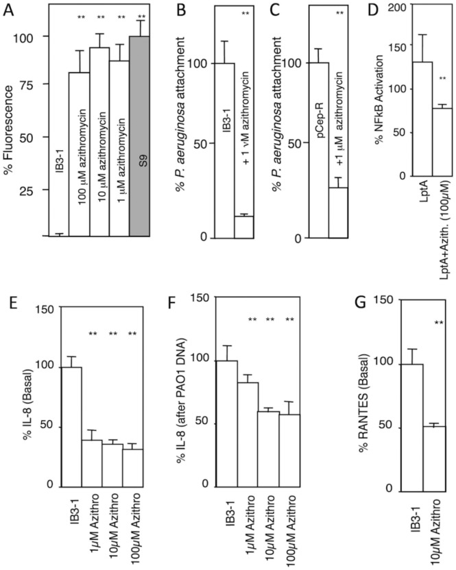
A. Cholera toxin (CTB-FITC fluorescence; used as aGM1 probe) binding to CF and normal cell monolayers grown for 14 days post-confluency and treated or not treated with azithomycin (48 h). IB3–1, CF cell line; S9, CFTR-ciorrecetd IB3–1 cells. B and C. P. aeruginosa (MOI of 1:200) attachment to IB3–1 (CF) or pCepR-16HBE (normal human bronchial epithelial cells rendered CF by expression of the R domain of CFTR) monolayers pretreated or not treated with AZT (the drug was absent from the adhesion assay). D. AZT reduces NFκB activation (luciferase reporter) in CF cells (IB3–1) in response to P. aeruginosa TLR2 ligand derived from lipoprotein LptA. E and F, AZT reduces basal IL-8 secretion and IL-8 induced by treatment with P. aeruginosa DNA. G, AZT reduces basal RANTES production by CF (IB3–1) cells. ** P (ANOVA; n=6) < 0.05.
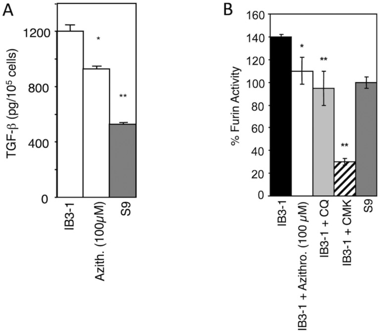
A. AZT effect on TGF-β secretion by CF (IB3–1) cells. B. Furin levels were corrected in full medium with AZT or 0.1 mM chloroquine (CQ). 50 μM furin inhibitor CMK was used as a measure of maximum furin inhibition. * P < 0.01, ** P < 0.05 (ANOVA, n=6).
References
Publication types
Grants and funding
LinkOut - more resources
Full Text Sources
Other Literature Sources