[Preprint]. 2020 Jul 13:2020.03.07.982264.
doi: 10.1101/2020.03.07.982264.

Type I interferon susceptibility distinguishes SARS-CoV-2 from SARS-CoV

Affiliations

Type I interferon susceptibility distinguishes SARS-CoV-2 from SARS-CoV

Kumari G Lokugamage et al. bioRxiv. .

Update in

Abstract

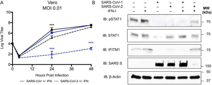
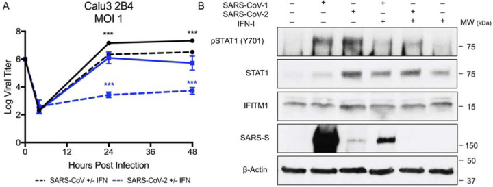
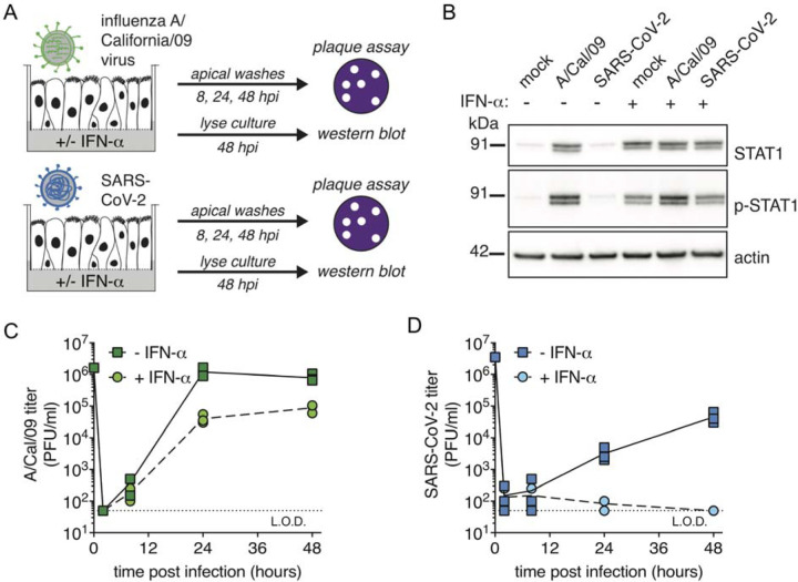
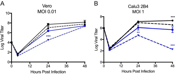
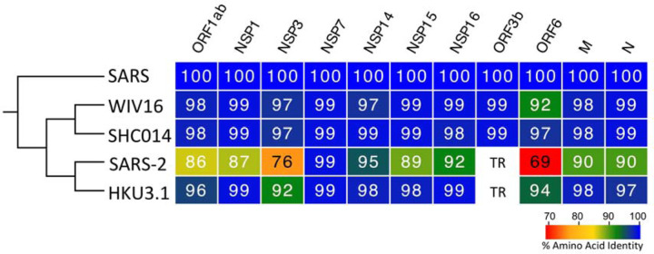
SARS-CoV-2, a novel coronavirus (CoV) that causes COVID-19, has recently emerged causing an ongoing outbreak of viral pneumonia around the world. While distinct from SARS-CoV, both group 2B CoVs share similar genome organization, origins to bat CoVs, and an arsenal of immune antagonists. In this report, we evaluate type-I interferon (IFN-I) sensitivity of SARS-CoV-2 relative to the original SARS-CoV. Our results indicate that while SARS-CoV-2 maintains similar viral replication to SARS-CoV, the novel CoV is much more sensitive to IFN-I. In Vero and in Calu3 cells, SARS-CoV-2 is substantially attenuated in the context of IFN-I pretreatment, while SARS-CoV is not. In line with these findings, SARS-CoV-2 fails to counteract phosphorylation of STAT1 and expression of ISG proteins, while SARS-CoV is able to suppress both. Comparing SARS-CoV-2 and influenza A virus in human airway epithelial cultures (HAEC), we observe the absence of IFN-I stimulation by SARS-CoV-2 alone, but detect failure to counteract STAT1 phosphorylation upon IFN-I pretreatment resulting in near ablation of SARS-CoV-2 infection. Next, we evaluated IFN-I treatment post infection and found SARS-CoV-2 was sensitive even after establishing infection. Finally, we examined homology between SARS-CoV and SARS-CoV-2 in viral proteins shown to be interferon antagonists. The absence of an equivalent open reading frame (ORF) 3b and changes to ORF6 suggest the two key IFN-I antagonists may not maintain equivalent function in SARS-CoV-2. Together, the results identify key differences in susceptibility to IFN-I responses between SARS-CoV and SARS-CoV-2 that may help inform disease progression, treatment options, and animal model development.

Keywords: 2019-nCoV; COVID-19; Coronavirus; IFN; SARS-CoV; SARS-CoV-2; type I interferon.


Figures

Figure 1.
Figure 1.. SARS-CoV-2 sensitive to type I IFN pretreatment.
A) Vero E6 cells were treated with 1000 U/mL recombinant type I (hashed line) IFN or mock (solid line) for 18 hours prior to infection. Cells were subsequently infected with either SARS-CoV WT (black) or SARS-CoV-2 (blue) at an MOI of 0.01 as described above. Each point on the line graph represents the group mean, N ≥ 3. All error bars represent SD. The two tailed students t-test was used to determine P-values: *** P < 0.001. B) Vero cell protein lysates from IFN-I treated and untreated cells were probed 48 hours post infection by Western blotting for phosphorylated STAT1 (Y701), STAT1, IFITM1, SARS spike, and Actin.
Figure 2.
Figure 2.. SARS-CoV-2 attenuated and IFN-I sensitive in Calu3 respiratory cells.
A) Calu3 2B4 cells were treated with 1000 U/mL recombinant type I (hashed line) IFN or mock (solid line) for 18 hours prior to infection. Cells were subsequently infected with either SARS-CoV WT (black) or SARS-CoV-2 (blue) at an MOI of 1. Each point on the line graph represents the group mean, N ≥ 3. All error bars represent SD. The two tailed students t-test was used to determine P-values: *** P < 0.001. B) Calu3 cell protein lysates from IFN-I treated and untreated cells were probed 48 hours post infection by Western blotting for phosphorylated STAT1 (Y701), STAT1, IFITM1, SARS spike, and Actin.
Figure 3.
Figure 3.. Differential IFN-I sensitivity and pSTAT1 phosphorylation following SARS-CoV-2 or influenza A virus on polarized human airway epithelial cultures (HAEC).
A) HAEC were pretreated with 1000 U/mL IFN-α basolaterally for 2 hours prior to and during infection. Cultures were then infected apically with influenza A/California/09 H1N1 virus or SARS-CoV-2. Apical washes were collected at the indicated times and progeny titers determined by plaque assay on MDCK cells (influenza virus) or Vero E6 cells (SARS-CoV-2). At the 48 h endpoint, cultures were lysed for western blot analysis. B) Western for total STAT1 or phospho-STAT1 at 48 hpi, and actin as loading control. C) Influenza A virus titers by plaque assay on MDCK cells. 0 h, virus inoculate; 2 h, virus in third apical wash; 8, 24, 48 h, virus in apical washes at these time points. D) SARS-CoV-2 titers by plaque assay on Vero E6 cells. 0 h, virus inoculate; 2 h, virus in second apical wash; 8, 24, 48 h, virus in apical washes at these time points.
Figure 4,
Figure 4,. SARS-CoV-2 impacted by post IFN-I treatment.
A-B) Vero E6 and Calu3 2B4 cells were infected with either SARS-CoV WT (black) or SARS-CoV-2 (blue) at an MOI 0.01 (A, Vero cells) or MOI 1 (B, Calu3 cells). Cells were subsequently treated with 1000 U/mL recombinant type I IFN (hashed line) or mock (solid line) for 4 hours following infection. Each point on the line graph represents the group mean, N ≥ 3. All error bars represent SD. The two tailed student’s t-test was used to determine P-values: **P <0.01 *** P < 0.001.
Figure 5,
Figure 5,. Conservation of SARS-CoV IFN antagonists.
Viral protein sequences of the indicated viruses were aligned according to the bounds of the SARS-CoV open reading frames for each viral protein. Sequence identities were extracted from the alignments for each viral protein, and a heat map of percent sequence identity was constructed using EvolView (www.evolgenius.info/evolview) with SARS-CoV as the reference sequence. TR = truncated protein.

References

    1. Gralinski LE, Menachery VD. 2020. Return of the Coronavirus: 2019-nCoV. Viruses 12. - PMC - PubMed
    1. Gorbalenya AE, Baker SC, Baric RS, de Groot RJ, Drosten C, Gulyaeva AA, Haagmans BL, Lauber C, Leontovich AM, Neuman BW, Penzar D, Perlman S, Poon LLM, Samborskiy DV, Sidorov IA, Sola I, Ziebuhr J, Coronaviridae Study Group of the International Committee on Taxonomy of V. 2020. The species Severe acute respiratory syndrome-related coronavirus: classifying 2019-nCoV and naming it SARS-CoV-2. Nature Microbiology doi:10.1038/s41564-020-0695-z - DOI - PMC - PubMed
    1. Zhu N, Zhang D, Wang W, Li X, Yang B, Song J, Zhao X, Huang B, Shi W, Lu R, Niu P, Zhan F, Ma X, Wang D, Xu W, Wu G, Gao GF, Tan W, China Novel Coronavirus I, Research T. 2020. A Novel Coronavirus from Patients with Pneumonia in China, 2019. N Engl J Med 382:727–733. - PMC - PubMed
    1. Huang C, Wang Y, Li X, Ren L, Zhao J, Hu Y, Zhang L, Fan G, Xu J, Gu X, Cheng Z, Yu T, Xia J, Wei Y, Wu W, Xie X, Yin W, Li H, Liu M, Xiao Y, Gao H, Guo L, Xie J, Wang G, Jiang R, Gao Z, Jin Q, Wang J, Cao B. 2020. Clinical features of patients infected with 2019 novel coronavirus in Wuhan, China. Lancet 395:497–506. - PMC - PubMed
    1. Wu Z, McGoogan JM. 2020. Characteristics of and Important Lessons From the Coronavirus Disease 2019 (COVID-19) Outbreak in China: Summary of a Report of 72314 Cases From the Chinese Center for Disease Control and Prevention. JAMA doi:10.1001/jama.2020.2648 - DOI - PubMed

Publication types