[Preprint]. 2020 May 18:2020.05.18.102087.
doi: 10.1101/2020.05.18.102087.

Controlling the SARS-CoV-2 Spike Glycoprotein Conformation

Affiliations

Controlling the SARS-CoV-2 Spike Glycoprotein Conformation

Rory Henderson et al. bioRxiv. .

Update in

  • Controlling the SARS-CoV-2 spike glycoprotein conformation.
    Henderson R, Edwards RJ, Mansouri K, Janowska K, Stalls V, Gobeil SMC, Kopp M, Li D, Parks R, Hsu AL, Borgnia MJ, Haynes BF, Acharya P. Henderson R, et al. Nat Struct Mol Biol. 2020 Oct;27(10):925-933. doi: 10.1038/s41594-020-0479-4. Epub 2020 Jul 22. Nat Struct Mol Biol. 2020. PMID: 32699321 Free PMC article.

Abstract

The coronavirus (CoV) viral host cell fusion spike (S) protein is the primary immunogenic target for virus neutralization and the current focus of many vaccine design efforts. The highly flexible S-protein, with its mobile domains, presents a moving target to the immune system. Here, to better understand S-protein mobility, we implemented a structure-based vector analysis of available β-CoV S-protein structures. We found that despite overall similarity in domain organization, different β-CoV strains display distinct S-protein configurations. Based on this analysis, we developed two soluble ectodomain constructs in which the highly immunogenic and mobile receptor binding domain (RBD) is locked in either the all-RBDs 'down' position or is induced to display a previously unobserved in SARS-CoV-2 2-RBDs 'up' configuration. These results demonstrate that the conformation of the S-protein can be controlled via rational design and provide a framework for the development of engineered coronavirus spike proteins for vaccine applications.


Figures

Figure 1.
Figure 1.. Vector based analysis of the β-CoV S-protein demonstrates remarkable variability in S-protein conformation within ‘up’ and ‘down’ states between CoV strains.
A) Cartoon representations of the ‘down’ (upper left) and ‘up’ (upper right) state structures colored according to the specified domains (lower). B) A single protomer of the β-CoV S-protein with labeled domains. C) A simplified diagram of the β-CoV S-protein depicting the centroids and vectors connecting them with the determine angles (θ) and dihedrals (ϕ) labeled. D) The SARS-2 (left; red) and MERS (right; blue) structures each with a single protomer depicted in a cartoon representation and the remaining two in a surface representation. E) Principal components analysis of the SARS, SARS-2, and MERS protomers including measures between S1 and S2 domains. F) Principal components analysis of the SARS, SARS-2, MERS, HKU1, and Murine CoV protomers including measures only between S1 domains.
Figure 2.
Figure 2.. Vector based analysis of the CoV S-protein demonstrates remarkable variability in S-protein conformation within ‘up’ and ‘down’ states between CoV strains.
A) Angle between the subdomain 1 to subdomain 2 vector and the subdomain 1 to RBD vector. B) Dihedral about the subdomain 1 to RBD vector. C) Angle between the RBD to subdomain 1 vector and the RBD to RBD helix vector. D) Dihedral about the subdomain 2 to subdomain 1 vector. E) Angle between the NTD’ to NTD vector and the NTD to NTD sheet motif vector. F) Dihedral about the NTD to NTD’ vector. G) Angle between the NTD’ to subdomain 2 vector and the NTD’ to NTD vector. H) Angle between the subdomain 2 to NTD’ vector and the subdomain 2 to subdomain 1 vector. I) Diagram of the domains and relevant angles and dihedrals for S1 J) Cartoon representation of one protomer’s S1 domains in the ‘down’ state overlaid with a ribbon representation of the ‘up’ state colored according to (I). Black (‘down’ state) and grey (‘up’ state) spheres represent domain centroids with lines connecting representing the vectors. Adjacent protomers represented as transparent surfaces.
Figure 3.
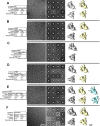
Figure 3.. Negative stain electron microscopy analysis of S-protein constructs.
A) Data tables, indicating construct names, mutations, observed classes, number and percent of particles per class and final resolution (gold-standard Fourier-shell correlation, 0.143 level). B) Raw micrographs. C) Representative 2D class averages. D) 3D reconstructions of 3-RBD-down classes, shown in top view, looking down the S-protein 3-fold axis on the left and tilted view on the right. E) 3D reconstructions of 1-RBD-up classes. Up-RBD is marked with an asterisk. F) 3D reconstruction of 2-RBD-up class. Density for up-RBDs is weak, indicated by asterisks. Receptor binding domains and N-terminal domains of first structure marked with R and N, respectively.
Figure 4.
Figure 4.. Cryo-EM dataset reveals differential stabilization of the S-protein in the mutant ectodomain constructs.
A) The S-protein spike highlighting the two regions of interest for structure and computation-based design. B) (top) The rS2d RBD to S2 locked structure displaying only the all RBD down state. (bottom) C) (top) The u1S2q SD1 to S2 mutated structure displaying the all RBD ‘down’ state, the 1-RBD ‘up’ state, and, for the first time in the SARS-2 S ectodomain, the 2-RBD ‘up’ state.
Figure 5.
Figure 5.. Cryo-EM structures of the “down” state in the r2S2d and u1S2q constructs reveal differential stabilization of domain positions.
A) Alignment between the trimers of the designed disulfide linked rS2d (dark blue) mutant structure and the u1S2q (green). B) (left) Alignment between single protomers of the designed disulfide linked rS2d(dark blue) mutant structure and the u1S2q(green). (right) Zoomed in view of SD1 in both constructs demonstrating the shift in the subdomain with the 4 mutants. C) Structure and cryo-EM map depicting the RBD to S2 bridging density between the introduced cysteine residues. D) Structure and cryo-EM map depicting the SD1 and S2 mutations.
Figure 6.
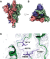
Figure 6.. High-resolution structure of the u1S2q 1 RBD ‘up’ state reveals increasing relaxation of the triggered RBDs toward the unmutated structure.
A) Cryo-EM reconstruction colored by chain. The RBD in the ‘up’ position is marked with an asterisk; side (left) and top (right) views. B) Zoomed-in view showing the mutated residues. C) (top) Structure of the ‘up’ state RBD coupled ‘down’ state RBD (green) highlighting the shifted subdomain 1 to NTD’ position relative to the unmutated position (blue). (middle) Structure of the uncoupled ‘down’ state RBD (green) highlighting the moderately shifted subdomain 1 to NTD’ position relative to the unmutated position (blue). (bottom) Structure of the ‘up’ state RBD (green) highlighting the close alignment of subdomain 1 and the NTD’ regions to the unmutated position (blue).
Figure 7.
Figure 7.. Structure of the u1S2q 2 RBD ‘up’ state indicates modest differences between the 1 RBD ‘up’ state’s subdomain arrangement.
A) Cryo-EM reconstruction colored by chain. The RBDs in the ‘up’ position are marked with asterisks; side (left) and top (right) views. B) Cryo-EM map structural alignment top view. C) Structure (green) and cryo-EM map depicting the mutated residue dispositions. The unmutated ‘up’ state protomer alignment is depicted in ribbons (blue).

References

    1. Hoffmann M. et al. SARS-CoV-2 Cell Entry Depends on ACE2 and TMPRSS2 and Is Blocked by a Clinically Proven Protease Inhibitor. Cell 181, 271–280 e278, doi: 10.1016/j.cell.2020.02.052 (2020). - DOI - PMC - PubMed
    1. Kirchdoerfer R. N. et al. Pre-fusion structure of a human coronavirus spike protein. Nature 531, 118–121, doi: 10.1038/nature17200 (2016). - DOI - PMC - PubMed
    1. Wrapp D. et al. Cryo-EM structure of the 2019-nCoV spike in the prefusion conformation. Science 367, 1260–1263, doi: 10.1126/science.abb2507 (2020). - DOI - PMC - PubMed
    1. Yuan Y. et al. Cryo-EM structures of MERS-CoV and SARS-CoV spike glycoproteins reveal the dynamic receptor binding domains. Nature Communications 8, 15092, doi: 10.1038/ncomms15092 (2017). - DOI - PMC - PubMed
    1. Gui M. et al. Cryo-electron microscopy structures of the SARS-CoV spike glycoprotein reveal a prerequisite conformational state for receptor binding. Cell Research 27, 119–129, doi: 10.1038/cr.2016.152 (2017). - DOI - PMC - PubMed

Publication types