[Preprint]. 2020 May 15:2020.05.11.088674.
doi: 10.1101/2020.05.11.088674.

Rapid isolation of potent SARS-CoV-2 neutralizing antibodies and protection in a small animal model

Affiliations

Rapid isolation of potent SARS-CoV-2 neutralizing antibodies and protection in a small animal model

Thomas F Rogers et al. bioRxiv. .

Update in

  • Isolation of potent SARS-CoV-2 neutralizing antibodies and protection from disease in a small animal model.
    Rogers TF, Zhao F, Huang D, Beutler N, Burns A, He WT, Limbo O, Smith C, Song G, Woehl J, Yang L, Abbott RK, Callaghan S, Garcia E, Hurtado J, Parren M, Peng L, Ramirez S, Ricketts J, Ricciardi MJ, Rawlings SA, Wu NC, Yuan M, Smith DM, Nemazee D, Teijaro JR, Voss JE, Wilson IA, Andrabi R, Briney B, Landais E, Sok D, Jardine JG, Burton DR. Rogers TF, et al. Science. 2020 Aug 21;369(6506):956-963. doi: 10.1126/science.abc7520. Epub 2020 Jun 15. Science. 2020. PMID: 32540903 Free PMC article.

Abstract

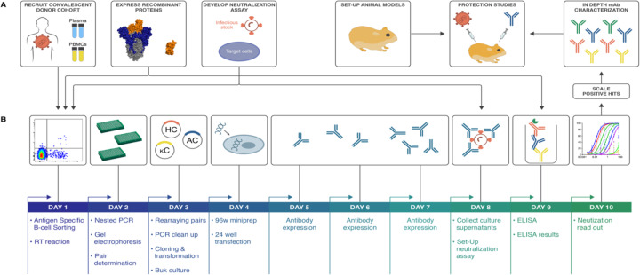
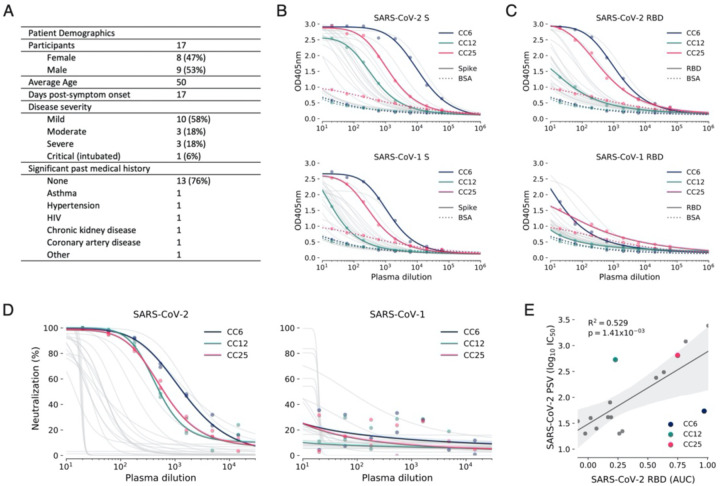
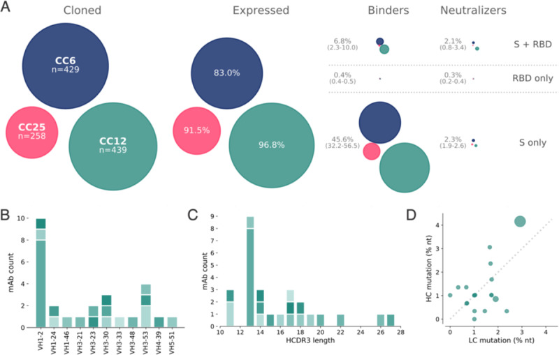
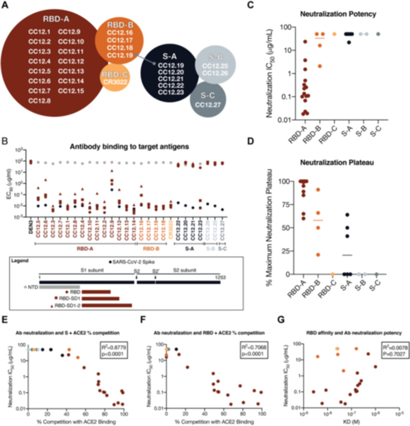
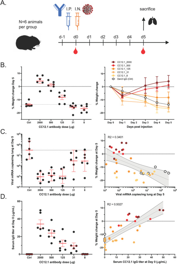
The development of countermeasures to prevent and treat COVID-19 is a global health priority. In under 7 weeks, we enrolled a cohort of SARS-CoV-2-recovered participants, developed neutralization assays to interrogate serum and monoclonal antibody responses, adapted our high throughput antibody isolation, production and characterization pipeline to rapidly screen over 1000 antigen-specific antibodies, and established an animal model to test protection. We report multiple highly potent neutralizing antibodies (nAbs) and show that passive transfer of a nAb provides protection against high-dose SARS-CoV-2 challenge in Syrian hamsters. The study suggests a role for nAbs in prophylaxis, and potentially therapy, of COVID-19. The nAbs define protective epitopes to guide vaccine design.


Figures

Figure 1.
Figure 1.. SARS-CoV-2 neutralizing antibody isolation strategy.
(A) A natural infection cohort was established to collect plasma and PBMCs samples from individuals who recovered from COVID-19. In parallel, functional assays were developed to rapidly screen all plasma samples for SARS-CoV-2neutralizing activity. SARS-CoV-2 recombinant surface proteins were also produced to use as baits in single memory B-cell sorting and downstream functional characterization of isolated mAbs. Finally, a hamster animal model was set-up to evaluate mAb passive transfer protection. (B) The standard mAb isolation pipeline was optimized to allow high-throughput amplification, cloning, expression and functional screening of hundreds of unpurified Ab heavy and light chain pairs isolated from each of several selected neutralizers in only 10 days. Selected pairs were scaled-up to purify IgG for validation and characterization experiments. The most potent neutralizing mAb was selected to evaluate protection in the Syrian hamster model.
Figure 2.
Figure 2.. COVID-19 cohort functional screening.
(A) Demographics of the UCSD COVID-19 cohort (CC) participants. CC plasma were tested for binding to SARS-CoV-1 and SARS-CoV-2 S protein (B) and RBD subunit (C) by ELISA. Background binding of plasma to BSA-coated plates is represented by a dashed line. (D) Plasma were also tested for neutralization of pseudotyped (PSV) SARS-CoV-1 and SARS-CoV-2 virions. (E) Correlation between PSV SARS-CoV-2 neutralization and RBD subunit ELISA binding area-under-the-curve (AUC). AUC was computed using Simpson’s rule. The 95% confidence interval of the regression line is shown in light grey and was estimated by performing 1,000 bootstrap re-samplings. R2 and p values of the regression are also indicated. CC participants from whom mAbs were isolated are specifically highlighted in dark blue (CC6), pine green (CC12) and hot pink (CC25).
Figure 3.
Figure 3.. Antibody isolation and functional screening for SARS-CoV neutralization and antigen binding.
(A) Antibody downselection process from 3 donors, presented as bubble plots. The areas of the bubbles for each donor are sized based on the number of antibodies that were cloned and transfected, then scaled according to the number that were positive in subsequent assays. All antibodies that expressed at measurable levels were tested for binding to S protein and RBD to determine their specificity, and then screened for neutralization. (B) VH gene distribution of downselected mAbs. Antibodies are colored by their respective clonal lineages. (C) Heavy chain CDR3 lengths of downselected mAbs. Antibodies are colored by their respective clonal lineages. (D) Mutation frequency of downselected mAb lineages. Bubble position represents the mean mutation frequency for each lineage, with bubble area proportional to the lineage size.
Figure 4.
Figure 4.. Antibody functional activity by epitope specificities.
Monoclonal antibody epitope binning was completed using RBD and SARS-CoV-2 S protein as target antigens. (A) A total of three non-competing epitopes for RBD (RBD-A, RBD-B, and RBD-C) and three non-competing epitopes for S (S-A, S-B, and S-C) were identified. (B) MAbs were evaluated for binding to different target antigens (S, N-terminal domain (NTD), RBD, RBD-SD1, and RBD-SD1–2) by ELISA and apparent EC50s are reported in μg/ml. (C) MAbs were evaluated for neutralization on SARS-CoV-2 pseudovirus and HeLa-ACE2 target cells. Antibodies are grouped according to epitope specificities and neutralization IC50 values are reported in μg/ml. (D) The maximum plateaus of neutralization (MPN) are reported for each mAb and grouped by epitope specificity. MAbs were mixed with (E) RBD or (F) S protein and measured for binding to HeLa-ACE2 target cells as a measure of competition to the cell surface ACE-2 receptor. (G) Monoclonal antibody neutralization potencies (IC50, μg/ml) are plotted compared to dissociation constants (KD, M) measured by surface plasmon resonance (SPR) to RBD target antigen.
Figure 5.
Figure 5.. A potent SARS-CoV-2 RBD-specific neutralizing mAb protects against disease progression and lung viral burden in Syrian hamsters.
(A) SARS-CoV-2-specific human neutralizing mAb CC12.1 isolated from natural infection was injected intraperitoneally into Syrian hamsters at a starting dose of 2 mg/animal (on average 16.5 mg/kg) and subsequent serial 4-fold dilutions. Control animals received 2 mg of a dengue-specific human IgG1 (Den3). Each group of 6 animals were challenged intranasally 12h post-infusion with 1X10⁶ PFU of SARS-CoV-2. Serum was collected at the time of challenge (Day 0) and Day 5, and their weight monitored as an indicator of disease progression. On day 5, lung tissue was collected for viral burden assessment. (B) Percentage weight change was calculated from day 0 for all animals at all time points. (C) Viral load as assessed by Q-PCR from lung tissue at day 5. (D) Serum titers of the passively administered mAb, as assessed by ELISA at the time of challenge (12h after i.p administration). Correlation analyses with 95% confidence intervals indicated in grey shade. R2 values are also indicated.

References

    1. The IMpact-RSV Study Group. Palivizumab, a Humanized Respiratory Syncytial Virus Monoclonal Antibody, Reduces Hospitalization From Respiratory Syncytial Virus Infection in High-risk Infants. Pediatrics. 1998;102: 531–537. - PubMed
    1. Corti D, Misasi J, Mulangu S, Stanley DA, Kanekiyo M, Wollen S, et al. Protective monotherapy against lethal Ebola virus infection by a potently neutralizing antibody. Science. 2016;351: 1339–1342. - PubMed
    1. Misasi J, Gilman MSA, Kanekiyo M, Gui M, Cagigi A, Mulangu S, et al. Structural and molecular basis for Ebola virus neutralization by protective human antibodies. Science. 2016;351: 1343–1346. - PMC - PubMed
    1. Walker LM, Burton DR. Passive immunotherapy of viral infections: “super-antibodies” enter the fray. Nat Rev Immunol. 2018;18: 297–308. - PMC - PubMed
    1. Giroglou T, Cinatl J Jr, Rabenau H, Drosten C, Schwalbe H, Doerr HW, et al. Retroviral vectors pseudotyped with severe acute respiratory syndrome coronavirus S protein. J Virol. 2004;78: 9007–9015. - PMC - PubMed

Publication types