[Preprint]. 2020 Apr 16:2020.03.17.20037713.
doi: 10.1101/2020.03.17.20037713.

A serological assay to detect SARS-CoV-2 seroconversion in humans

Affiliations

A serological assay to detect SARS-CoV-2 seroconversion in humans

Fatima Amanat et al. medRxiv. .

Update in

  • A serological assay to detect SARS-CoV-2 seroconversion in humans.
    Amanat F, Stadlbauer D, Strohmeier S, Nguyen THO, Chromikova V, McMahon M, Jiang K, Arunkumar GA, Jurczyszak D, Polanco J, Bermudez-Gonzalez M, Kleiner G, Aydillo T, Miorin L, Fierer DS, Lugo LA, Kojic EM, Stoever J, Liu STH, Cunningham-Rundles C, Felgner PL, Moran T, García-Sastre A, Caplivski D, Cheng AC, Kedzierska K, Vapalahti O, Hepojoki JM, Simon V, Krammer F. Amanat F, et al. Nat Med. 2020 Jul;26(7):1033-1036. doi: 10.1038/s41591-020-0913-5. Epub 2020 May 12. Nat Med. 2020. PMID: 32398876 Free PMC article.

Abstract

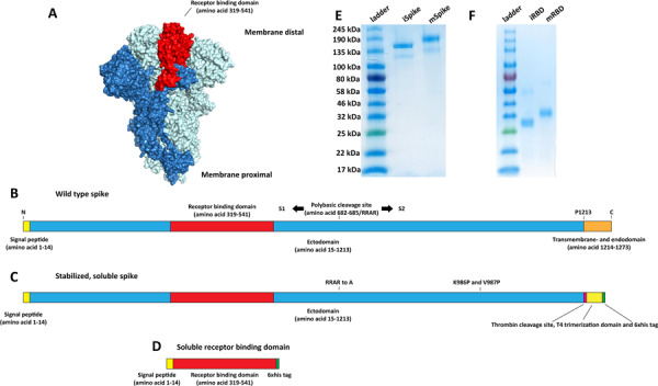
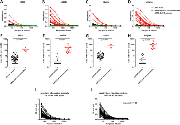
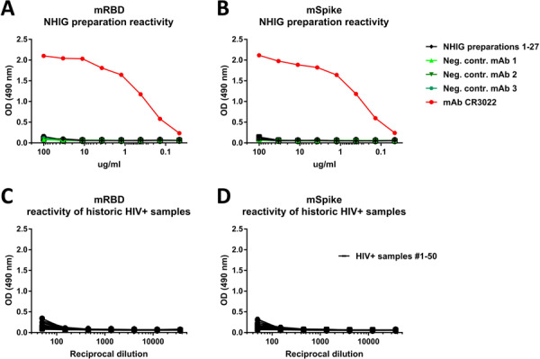
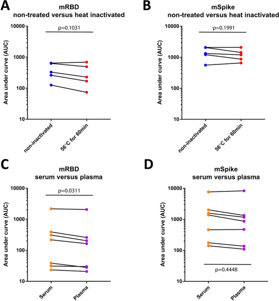
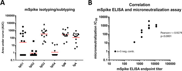
SARS-Cov-2 (severe acute respiratory disease coronavirus 2), which causes Coronavirus Disease 2019 (COVID19) was first detected in China in late 2019 and has since then caused a global pandemic. While molecular assays to directly detect the viral genetic material are available for the diagnosis of acute infection, we currently lack serological assays suitable to specifically detect SARS-CoV-2 antibodies. Here we describe serological enzyme-linked immunosorbent assays (ELISA) that we developed using recombinant antigens derived from the spike protein of SARS-CoV-2. Using negative control samples representing pre-COVID 19 background immunity in the general adult population as well as samples from COVID19 patients, we demonstrate that these assays are sensitive and specific, allowing for screening and identification of COVID19 seroconverters using human plasma/serum as early as two days post COVID19 symptoms onset. Importantly, these assays do not require handling of infectious virus, can be adjusted to detect different antibody types and are amendable to scaling. Such serological assays are of critical importance to determine seroprevalence in a given population, define previous exposure and identify highly reactive human donors for the generation of convalescent serum as therapeutic. Sensitive and specific identification of coronavirus SARS-Cov-2 antibody titers may, in the future, also support screening of health care workers to identify those who are already immune and can be deployed to care for infected patients minimizing the risk of viral spread to colleagues and other patients.


Conflict of interest statement

Conflict of interest Mount Sinai is in the process of licensing out assays based on the assays described here to commercial entities.

Figures

Figure 1:
Figure 1:. Constructs for recombinant protein expression.
A Visualization of the trimeric spike protein of SARS-CoV-2 based on PBD # 6VXX using Pymol. One monomer is colored in dark blue while the remaining two monomers are held in light blue. The receptor binding domain (RBD) of the dark blue trimer is highlighted in red. B Schematic of the wild type full length spike protein with signal peptide, ectodomain, receptor binding domain, furin cleavage site, S1, S2, and transmembrane and endodomain domain indicated. C Schematic of the soluble trimeric spike. The polybasic/furin cleavage site (RRAR) was replaced by a single A. The transmembrane and endodomain were replaced by a furin cleavage site, a T4 foldon tetramerization domain and a hexahistidine tag. Introduction of K986P and V987P has been shown to stabilize the trimer in the pre-fusion conformation. D Schematic of the soluble receptor binding domain construct. All constructs are to scale. E Reducing SDS PAGE of insect cell and mammalian cell derived soluble trimerized spike protein (iSpike and mSpike). F Reducing SDS PAGE of insect cell derived and mammalian cell derived recombinant receptor binding domain (iRBD and mRBD).
Figure 2:
Figure 2:. Reactivity of control and SARS-CoV-2 convalescent sera to different spike antigens.
A-D Reactivity to insect cell derived RBD (iRBD), mammalian cell derived RBD (mRBD), insect cell derived soluble spike protein (iSpike) and mammalian cell derived soluble spike protein (mSpike). Sera from SARS-CoV-2 infected individuals are shown in red. One sample, shown in green, is a convalescent serum sample post NL63 infection. E-F shows data from the same experiment but graphed as area under the curve (AUC) to get a better quantitative impression. The n for the control samples is 50 except for the iRBD where it is 59. Statistics were performed using an unpaired two-tailed student’s t-test in Graphpad Prism. I-G shows reactivity of the 50 negative control samples from A-F against spike protein from human coronaviruses 229E and NL63.
Figure 3:
Figure 3:. Human normal immunoglobulin preparations and historic sera from HIV+ patients do not react with the SAR-CoV-2 spike.
A-B Reactivity of 21 different pools of human normal immunoglobulin (HNIG) preparations (27 different vials) to mRBD and mSpike of SARS-CoV-2. MAb CR3022 was used as positive control, three different irrelevant human mAbs were used as negative control. C-D shows reactivity of historic samples from 50 HIV+ individuals to mRBD and mSpike of SARS-CoV-2. Both HNIG and serum samples from HIV+ donors were collected before the SARS-CoV-2 pandemic.
Figure 4:
Figure 4:. Effect of heat treatment and serum versus plasma on assay performance.
A-B Reactivity of paired non-treated serum and heat treated serum samples to mRBD and mSpike of SARS-CoV-2 (n=5). C-D Reactivity of paired serum and plasma samples to mRBD and mSpike of SARS-CoV-2 (n=7). Statistics were performed using a paired student’s t-test in Graphpad Prism.
Figure 5:
Figure 5:. Isotypes and subtypes of antibodies from COVID19 patients to the soluble spike protein and correlation between ELISA and microneutralization titer.
(A) Mammalian cell derived spike protein was used to study isotype/subclass distribution of antibodies (n=13 positive samples). (B) Correlation between ELISA titers and microneutralization titers (n=12, the three samples from negative control sera overlap and are displayed as single point). Statistics were performed using Pearson’s rank test in Graphpad Prism.

References

    1. Wu F., et al. A new coronavirus associated with human respiratory disease in China. Nature (2020). - PMC - PubMed
    1. Zhou P., et al. A pneumonia outbreak associated with a new coronavirus of probable bat origin. Nature (2020). - PMC - PubMed
    1. Gorbalenya A.E., et al. The species Severe acute respiratory syndrome-related coronavirus: classifying 2019-nCoV and naming it SARS-CoV-2. Nature Microbiology (2020). - PMC - PubMed
    1. Chu D.K.W., et al. Molecular Diagnosis of a Novel Coronavirus (2019-nCoV) Causing an Outbreak of Pneumonia. Clin Chem (2020). - PMC - PubMed
    1. Corman V.M., et al. Detection of 2019 novel coronavirus (2019-nCoV) by real-time RT-PCR. Euro Surveill 25(2020). - PMC - PubMed

Publication types