

The development and validation of a radiomic nomogram for the preoperative prediction of lung adenocarcinoma
- PMID: 32513144
- PMCID: PMC7278188
- DOI: 10.1186/s12885-020-07017-7
The development and validation of a radiomic nomogram for the preoperative prediction of lung adenocarcinoma
Abstract
Background: Accurate diagnosis of early lung cancer from small pulmonary nodules (SPN) is challenging in clinical setting. We aimed to develop a radiomic nomogram to differentiate lung adenocarcinoma from benign SPN.
Methods: This retrospective study included a total of 210 pathologically confirmed SPN (≤ 10 mm) from 197 patients, which were randomly divided into a training dataset (n = 147; malignant nodules, n = 94) and a validation dataset (n = 63; malignant nodules, n = 39). Radiomic features were extracted from the cancerous volumes of interest on contrast-enhanced CT images. The least absolute shrinkage and selection operator (LASSO) regression was used for data dimension reduction, feature selection, and radiomic signature building. Using multivariable logistic regression analysis, a radiomic nomogram was developed incorporating the radiomic signature and the conventional CT signs observed by radiologists. Discrimination and calibration of the radiomic nomogram were evaluated.
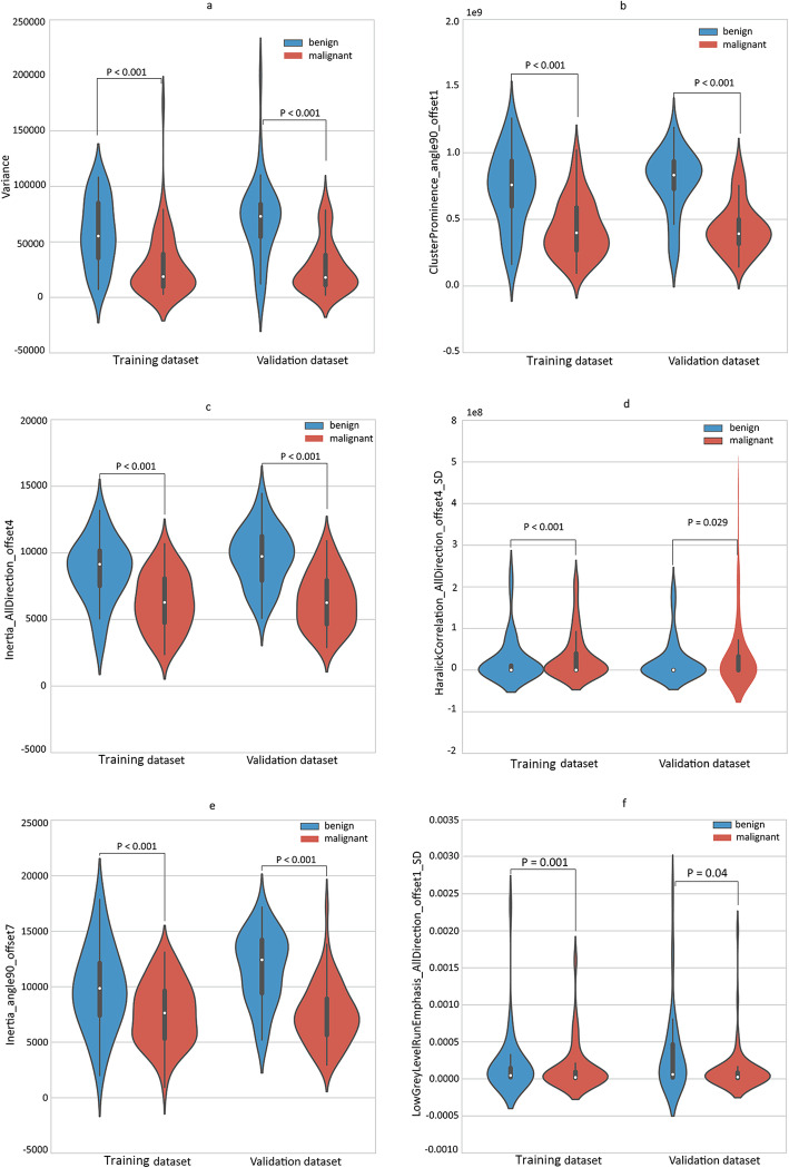
Results: The radiomic signature consisting of five radiomic features achieved an AUC of 0.853 (95% confidence interval [CI]: 0.735-0.970), accuracy of 81.0%, sensitivity of 82.9%, and specificity of 77.3%. The two conventional CT signs achieved an AUC of 0.833 (95% CI: 0.707-0.958), accuracy of 65.1%, sensitivity of 53.7%, and specificity of 86.4%. The radiomic nomogram incorporating the radiomic signature and conventional CT signs showed an improved AUC of 0.857 (95% CI: 0.723-0.991), accuracy of 84.1%, sensitivity of 85.4%, and specificity of 81.8%. The radiomic nomogram had good calibration power.
Conclusion: The radiomic nomogram might has the potential to be used as a non-invasive tool for individual prediction of SPN preoperatively. It might facilitate decision-making and improve the management of SPN in the clinical setting.
Keywords: Computed tomography; Diagnosis; Lung adenocarcinoma; Nomogram; Radiomics.
Conflict of interest statement
The authors declare that they have no competing interests.
Figures


Similar articles
-
A comparative study to evaluate CT-based semantic and radiomic features in preoperative diagnosis of invasive pulmonary adenocarcinomas manifesting as subsolid nodules.Sci Rep. 2021 Jan 18;11(1):66. doi: 10.1038/s41598-020-79690-4. Sci Rep. 2021. PMID: 33462251 Free PMC article.
-
Can peritumoral radiomics increase the efficiency of the prediction for lymph node metastasis in clinical stage T1 lung adenocarcinoma on CT?Eur Radiol. 2019 Nov;29(11):6049-6058. doi: 10.1007/s00330-019-06084-0. Epub 2019 Mar 18. Eur Radiol. 2019. PMID: 30887209
-
Preoperative diagnosis of malignant pulmonary nodules in lung cancer screening with a radiomics nomogram.Cancer Commun (Lond). 2020 Jan;40(1):16-24. doi: 10.1002/cac2.12002. Epub 2020 Mar 3. Cancer Commun (Lond). 2020. PMID: 32125097 Free PMC article.
-
Development of a nomogram-based model incorporating radiomic features from follow-up longitudinal lung CT images to distinguish invasive adenocarcinoma from benign lesions: a retrospective study.BMC Pulm Med. 2024 Oct 26;24(1):534. doi: 10.1186/s12890-024-03360-8. BMC Pulm Med. 2024. PMID: 39455958 Free PMC article.
-
Development and validation of a preoperative CT‑based radiomics nomogram to differentiate tuberculosis granulomas from lung adenocarcinomas: an external validation study.BMC Cancer. 2024 Jun 1;24(1):670. doi: 10.1186/s12885-024-12422-3. BMC Cancer. 2024. PMID: 38824514 Free PMC article.
Cited by
-
Feature selection methods and predictive models in CT lung cancer radiomics.J Appl Clin Med Phys. 2023 Jan;24(1):e13869. doi: 10.1002/acm2.13869. Epub 2022 Dec 17. J Appl Clin Med Phys. 2023. PMID: 36527376 Free PMC article. Review.
-
Statistical plots in oncologic imaging, a primer for neuroradiologists.Neuroradiol J. 2024 Aug;37(4):418-433. doi: 10.1177/19714009231193158. Epub 2023 Aug 2. Neuroradiol J. 2024. PMID: 37529843 Free PMC article. Review.
-
Nomogram integrating clinical-radiological and radiomics features for differentiating invasive from non-invasive pulmonary adenocarcinomas presenting as ground-glass nodules.Am J Cancer Res. 2025 Feb 15;15(2):797-810. doi: 10.62347/AOAN9966. eCollection 2025. Am J Cancer Res. 2025. PMID: 40084360 Free PMC article.
-
Identification of pulmonary adenocarcinoma and benign lesions in isolated solid lung nodules based on a nomogram of intranodal and perinodal CT radiomic features.Front Oncol. 2022 Sep 6;12:924055. doi: 10.3389/fonc.2022.924055. eCollection 2022. Front Oncol. 2022. PMID: 36147924 Free PMC article.
-
Differential diagnosis of benign and lung adenocarcinoma presenting as larger solid nodules and masses based on multiscale CT radiomics.PLoS One. 2024 Oct 4;19(10):e0309033. doi: 10.1371/journal.pone.0309033. eCollection 2024. PLoS One. 2024. PMID: 39365772 Free PMC article.
References
Publication types
MeSH terms
Grants and funding
LinkOut - more resources
Full Text Sources
Medical
