Review
. 2021 Oct 1;11(10):a038638.
doi: 10.1101/cshperspect.a038638.

Hemagglutinin Structure and Activities

Affiliations
Review

Hemagglutinin Structure and Activities

Steven J Gamblin et al. Cold Spring Harb Perspect Med. .

Abstract

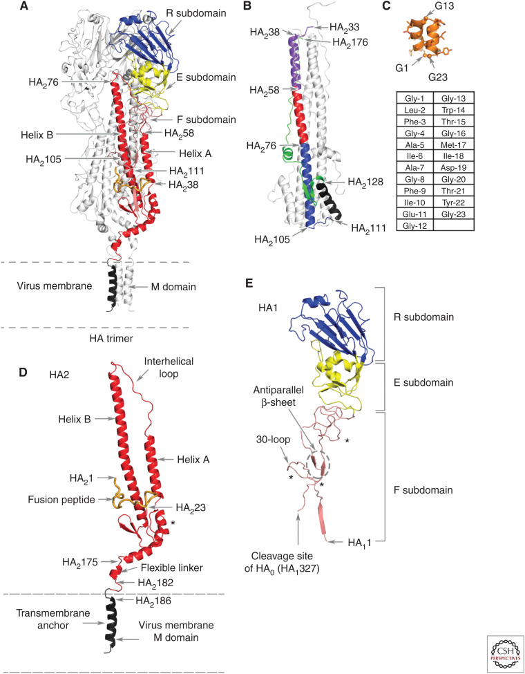
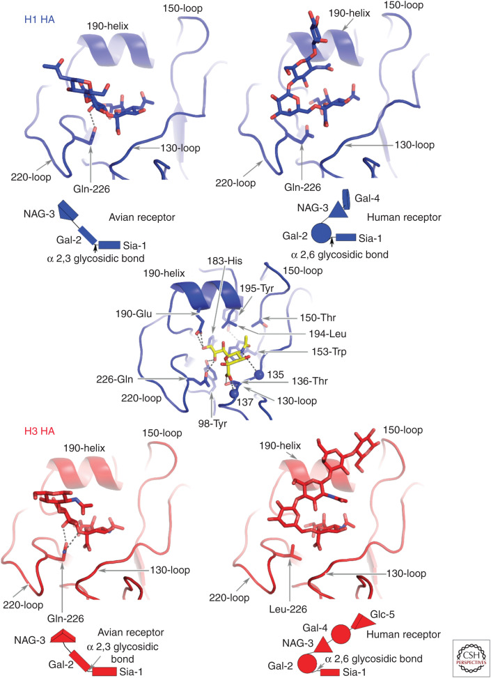
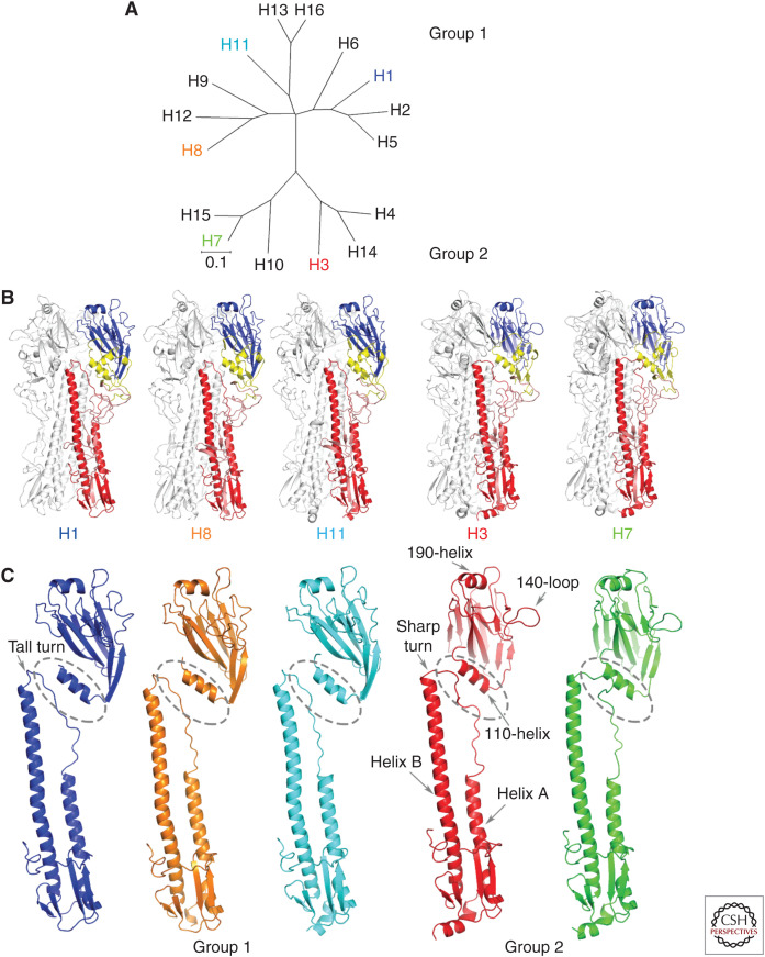
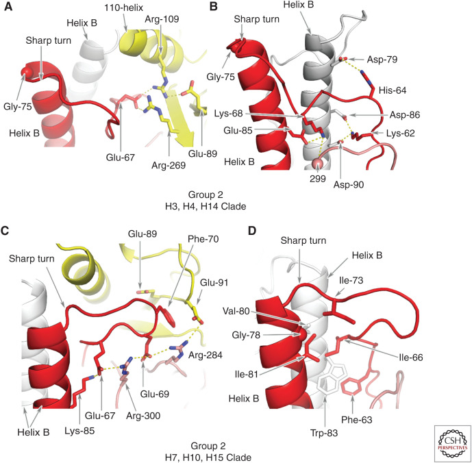
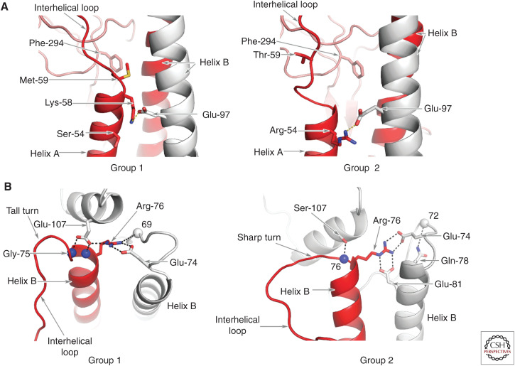
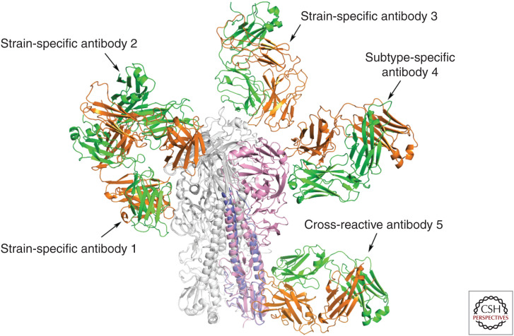
Hemagglutinins (HAs) are the receptor-binding and membrane fusion glycoproteins of influenza viruses. They recognize sialic acid-containing, cell-surface glycoconjugates as receptors but have limited affinity for them, and, as a consequence, virus attachment to cells requires their interaction with several virus HAs. Receptor-bound virus is transferred into endosomes where membrane fusion by HAs is activated at pH between 5 and 6.5, depending on the strain of virus. Fusion activity requires extensive rearrangements in HA conformation that include extrusion of a buried "fusion peptide" to connect with the endosomal membrane, form a bridge to the virus membrane, and eventually bring both membranes close together. In this review, we give an overview of the structures of the 16 genetically and antigenically distinct subtypes of influenza A HA in relation to these two functions in virus replication and in relation to recognition of HA by antibodies that neutralize infection.


Figures

Figure 1.
Figure 1.
The polypeptide and subdomain structure of hemagglutinin (HA). (A) An HA trimer of the H1 subtype with one subunit colored according to subdomains with the other two subunits in silver. From membrane-distal to membrane-anchor subdomains: the receptor-binding subdomain (blue), the vestigial esterase subdomain (yellow), the fusion subdomain (red), and the membrane anchor subdomain (black). (B) The fusion pH structure of an (X-31) H3 subtype HA2 expressed in Escherichia coli (Chen et al. 1999). Membrane fusion by influenza viruses is activated at acidic pH, in the case of X-31 at pH 5.6, and changes in the conformation of HA2 required for fusion result in the formation of this structure. The color code indicates the positions of the relocated helix-A (purple), the interhelical loop (red), and the coiled-coil component of helix B (blue) (which is unchanged from its conformation at neutral pH). The turn formed by residues 105–111 positions the carboxy-terminal region of helix B (black) and the carboxy-terminal components of the ectodomain, residues 129–176 (green), antiparallel to the central, 100 Å coiled-coil. Neither the fusion peptide nor the membrane anchor regions are components of this structure, but they would both be positioned at the same (top) end of the coiled-coil. (C) The structure of a synthetic 23-residue fusion peptide (orange), determined by nuclear magnetic resonance (NMR) (Lorieau et al. 2010), indicates that a monomer peptide forms a hairpin-like structure made of two antiparallel helices that turn around conserved glycine-13. Three of the seven conserved glycine residues in the peptide are indicated, as are the surface locations of large side-chain hydrophobic residues, eight of which are completely conserved. The sequence of the peptide is shown in the table below the structure. (D) The HA2 polypeptide from A showing (i) the interhelical loop, (ii) the 23-residue-long fusion peptide at the amino terminus (in orange), (iii) the flexible linker of the ectodomain to the membrane anchor, and (iv) the location of the single carbohydrate side chain (marked by *). (E) The HA1 polypeptide component of the colored monomer from A showing (i) the HA1 region of the fusion subdomain with its amino terminus near the virus membrane, (ii) the site of cleavage of the precursor HA0 that generates the carboxyl terminus of HA1 and the amino terminus of HA2, (iii) the antiparallel strands of the β-sheet and the extended chains in the fusion subdomain, and (iv) the locations of the carbohydrate side chains (marked by *).
Figure 2.
Figure 2.
Avian and human receptor analogs complexed with Group 1 and Group 2 HAs represented by H1 and H3 HAs. The saccharide composition of the receptors is shown schematically below each panel. The two types of receptors are distinguished by the α-2,3 (avian) and α-2,6 (human) linkages between sialic acid and the second saccharide, galactose. Different avian species also prefer oligosaccharides with different linkages between Gal-2 and GlcNAc-3. Wild ducks, for example, prefer Gal-2 b1,3-linked GlcNAc-3, whereas domesticated poultry prefer Gal-2 b1-4 GlcNAc-3-linked sialosides (Gambaryan et al. 2008). In the central panel sialic acid (yellow) forms hydrogen bonds between its carboxylate and the hydroxyl of Thr-136 and the peptide amide of residue 137, and between the nitrogen of the acetamido substituent and the carbonyl of residue 135. The methyl group of the acetamido makes hydrophobic contacts in a pocket formed of Leu-194, Trp-153, and Ile-155. The HAs of the pandemic viruses of 1957 (H2) and 1968 (H3) contained Leu-226 instead of the avian-specific Gln-226 of their proposed avian precursors. In the 1918, 1977, and 2009 H1 pandemic viruses, in contrast, Gln-226 was retained and substitutions Glu190Asp and Gly225Asn correlated with the receptor-binding specificity change to a preference for human receptor. In the characteristic avian receptor-binding motif (Ha et al. 2001) Gln-226 forms hydrogen bonds with the 4-hydroxyl group of Gal-2 and with the glycosidic oxygen of the α-2,3 linkage in the trans conformation. By contrast, the Leu-226 residue contacts carbon-6 of Gal-2 in the α-2,6- linkage in the cis configuration of the human receptor. The α-2,3-linked avian receptor analogs extend linearly from Gal-2 out of the site between the 190-helix and the 220-loop. Again, by contrast, the α-2,6-linked human receptor analog following Gal-2 bends back to different extents over sialic acid.
Figure 3.
Figure 3.
Sequence and structural similarity of HAs in the five clades. (A) Genetic relationships between HAs of the 16 subtypes of influenza A showing the three clades of Group 1 and the two clades of Group 2. (B) HA trimers and (C) HA monomers of the five clades compared. One representative of each clade is shown—H1, H8, and H11 of Group 1 and H3 and H7 of Group 2. Of note are the shorter helix A in Group 2 HAs; the sharp turn at the carboxyl terminus of the interhelical loop in Group 2 HAs and the tall turn in Group 1 HAs; the rotation of Group 1 membrane-distal subdomains relative to those of Group 2 HAs, noticeable from the orientation of the 190-helices at the membrane-distal tip of each subunit, and for the prominent 140-loop on the right side of Group 2 HAs; and the relative proximity of the 110-helix of the esterase subdomain to the interhelical loop in Group 2 HAs.
Figure 4.
Figure 4.
Group 2 HA interactions between residues in the interhelical loop, helix B, and the E subdomain. (A) In clade H3,4,14 HAs, an intrasubunit network of salt bridges between clade-specific HA2 Glu-67 and E subdomain residues HA1 Arg-109 and Arg-269 in H3 (Lys-269 in H14 and Asn-269 in H4.) HA1 Arg-109 also forms a salt bridge with HA1 Glu-89. (B) In clade H3,4,14 HAs, there are also interactions between Lys-62 of the loop and Asp-86 and Asp-90 of a neighboring helix B. There is an interaction between His-64 and Asp-79 of the same neighboring helix B, and between Lys-68 and Glu-85 of helix B of the same subunit. Lys-68 also interacts with the hydroxyl of residue HA1 299 of the F subdomain antiparallel β-sheet. (C) The sharp turn at the carboxyl terminus of the interhelical loop of clade H7,10,15 HAs is stabilized by intrasubunit contacts made by Phe-70 in a cavity formed by the aliphatic side chains of Glu-89 and Glu-91 of the E subdomain and Arg-284 of the F subdomain. Arg-284 also makes charged interactions with Glu-91 and Glu-69, and Arg-300 interacts with Glu-69 and Glu-67. (D) These contacts are augmented by hydrophobic interactions made by loop residues Phe-63, Ile-66, and Ile-73 (Val in H7 and H15) that pack against the nonpolar residues of helix B, Gly-78 and Ile-81, and Val-80 and Trp-83 of a neighboring helix B.
Figure 5.
Figure 5.
Group-specific structural features at the amino and carboxyl termini of the interhelical loop. (A) A comparison of the structures at the amino termini of the interhelical loops. The interactions of Group 1–conserved Met-59 with conserved Phe-294 (left panel) are shown by comparison with the lack of interaction with Group 2–conserved Thr-59 (right panel). In Group 2 HAs of clade 3,4,14 shown here, Group 2–conserved Glu-97 forms a salt bridge with Group 2–conserved Arg-54. In Group 2 clade H7,10,15, Arg-54 forms a salt bridge with conserved Glu-103 (not shown). (B) A comparison of the structures at the carboxyl termini of the interhelical loops indicating the tall turn adopted by Group 1 HAs, which is stabilized by a salt bridge between group-conserved Arg-76 and conserved Glu-107 of a neighboring subunit and by hydrogen bonds between conserved HA1 Glu-107 and the peptide amides of residues 75 and 76 and between Arg-76 and the carbonyl of residue 69. In Group 2 HAs clade 3,4,14, by contrast, the carboxyl terminus of the loop forms a sharp turn to the amino terminus of helix B, stabilized by salt bridges between group-conserved Arg-76 and conserved Glu-74 and clade-specific Glu-81 and a hydrogen bond between clade-specific Ser-107 and peptide amide 76 and between clade-specific Gln-78 and the peptide carbonyl of residue 72. None of these interactions are formed by clade H7,10,15 HAs.
Figure 6.
Figure 6.
Examples of the sites of binding of anti-HA monoclonal antibodies. The structures of complexes formed by Fabs of five monoclonal antibodies that were determined by X-ray crystallography illustrate the regions of HA recognized by antibodies that block infection. Antibodies 1, 2, and 3 bind in or near the receptor-binding site of HA. Antibodies 2 and 3 directly block receptor binding and are strain-specific. Other antibodies of this type with longer HCDR3 loops that penetrate the site can be cross-reactive within a subtype (Whittle et al. 2011; Ekiert et al. 2012). Antibody 1 binds ∼15 Å below the site and is less effective at blocking receptor binding. It may do so by projecting the Fc portion of the antibody over the receptor-binding site and sterically preventing receptor access. Antibody 4 binds at a similar distance from the site to antibody 1, but to the molecular surface behind the receptor-binding pocket. It also does not directly prevent receptor binding, but it blocks infection by all viruses in the H5 subtype. Cross-reactive antibody 5 binds near the fusion peptide and in vitro can block HA0 cleavage and also prevent the conformational change in HA required for fusion. It blocks infection by viruses of Group 1 and Group 2 as a result of immune cytolysis following recognition of the bound antibody by Fc receptor–bearing cells. Antibody 4 may block infection in a similar manner. Antibodies 1, 2, and 3 are from mice immunized with HA from the H3 subtype (Bizebard et al. 1995; Knossow et al. 2002). Antibodies 4 and 5 were isolated from immunized humans (Corti et al. 2011; Xiong et al. 2015; Kallewaard et al. 2016).

References

    1. Air GM. 1981. Sequence relationships among the hemagglutinin genes of 12 subtypes of influenza A virus. Proc Natl Acad Sci 78: 7639–7643. 10.1073/pnas.78.12.7639 - DOI - PMC - PubMed
    1. Baigent SJ, McCauley JW. 2001. Glycosylation of haemagglutinin and stalk-length of neuraminidase combine to regulate the growth of avian influenza viruses in tissue culture. Virus Res 79: 177–185. 10.1016/S0168-1702(01)00272-6 - DOI - PubMed
    1. Bangaru S, Lang S, Schotsaert M, Vanderven HA, Zhu X, Kose N, Bombardi R, Finn JA, Kent SJ, Gilchuk P, et al.2019. A site of vulnerability on the influenza virus hemagglutinin head domain trimer interface. Cell 177: 1136–1152.e18. 10.1016/j.cell.2019.04.011 - DOI - PMC - PubMed
    1. Benton DJ, Martin SR, Wharton SA, McCauley JW. 2015. Biophysical measurement of the balance of influenza A hemagglutinin and neuraminidase activities. J Biol Chem 290: 6516–6521. 10.1074/jbc.M114.622308 - DOI - PMC - PubMed
    1. Benton DJ, Nans A, Calder LJ, Turner J, Neu U, Lin YP, Ketelaars E, Kallewaard NL, Corti D, Lanzavecchia A, et al.2018. Influenza hemagglutinin membrane anchor. Proc Natl Acad Sci 115: 10112–10117. 10.1073/pnas.1810927115 - DOI - PMC - PubMed

LinkOut - more resources