. 2020 Jun 23;117(25):14412-14420.
doi: 10.1073/pnas.1920165117. Epub 2020 Jun 8.

Topoisomerase I-driven repair of UV-induced damage in NER-deficient cells

Affiliations

Topoisomerase I-driven repair of UV-induced damage in NER-deficient cells

Liton Kumar Saha et al. Proc Natl Acad Sci U S A. .

Abstract

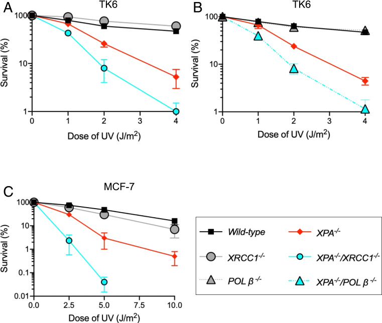
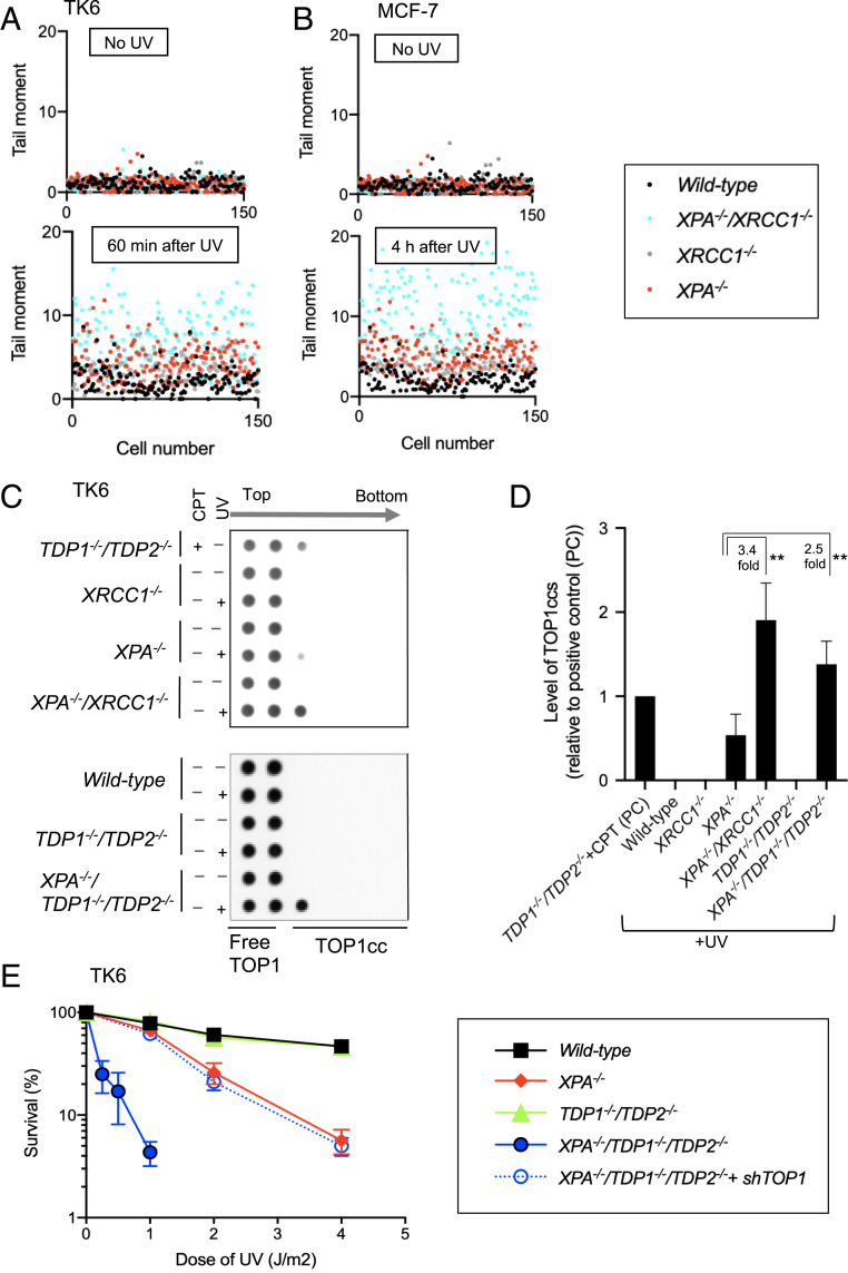
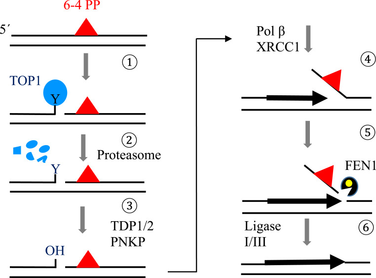
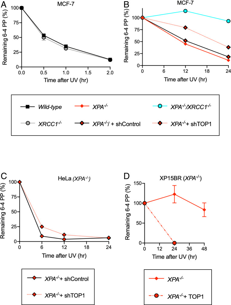
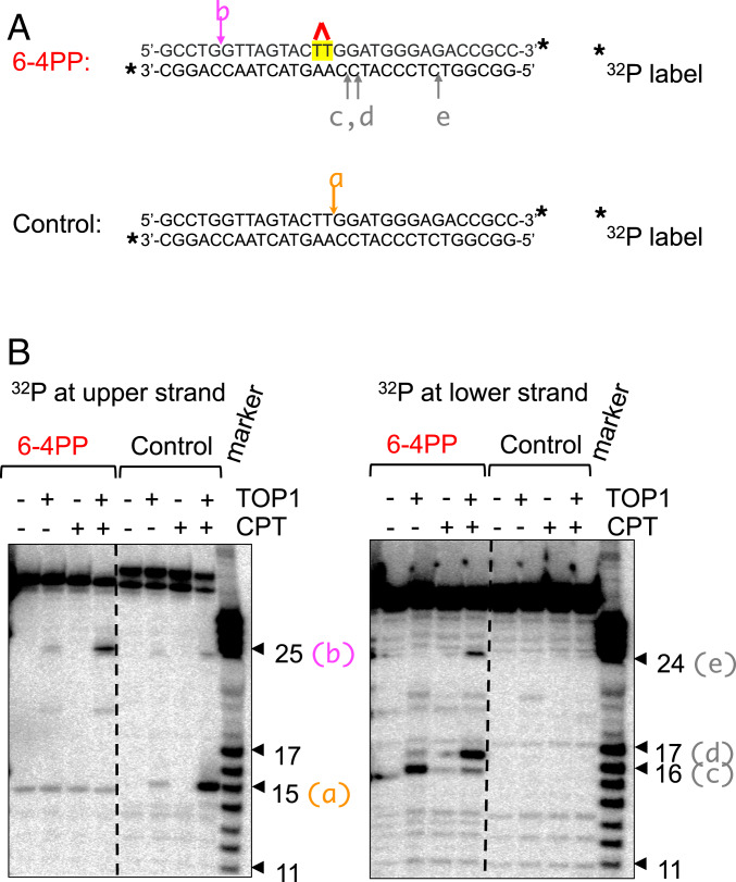
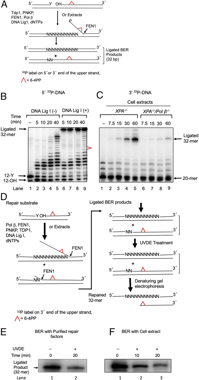
Nucleotide excision repair (NER) removes helix-destabilizing adducts including ultraviolet (UV) lesions, cyclobutane pyrimidine dimers (CPDs), and pyrimidine (6-4) pyrimidone photoproducts (6-4PPs). In comparison with CPDs, 6-4PPs have greater cytotoxicity and more strongly destabilizing properties of the DNA helix. It is generally believed that NER is the only DNA repair pathway that removes the UV lesions as evidenced by the previous data since no repair of UV lesions was detected in NER-deficient skin fibroblasts. Topoisomerase I (TOP1) constantly creates transient single-strand breaks (SSBs) releasing the torsional stress in genomic duplex DNA. Stalled TOP1-SSB complexes can form near DNA lesions including abasic sites and ribonucleotides embedded in chromosomal DNA. Here we show that base excision repair (BER) increases cellular tolerance to UV independently of NER in cancer cells. UV lesions irreversibly trap stable TOP1-SSB complexes near the UV damage in NER-deficient cells, and the resulting SSBs activate BER. Biochemical experiments show that 6-4PPs efficiently induce stable TOP1-SSB complexes, and the long-patch repair synthesis of BER removes 6-4PPs downstream of the SSB. Furthermore, NER-deficient cancer cell lines remove 6-4PPs within 24 h, but not CPDs, and the removal correlates with TOP1 expression. NER-deficient skin fibroblasts weakly express TOP1 and show no detectable repair of 6-4PPs. Remarkably, the ectopic expression of TOP1 in these fibroblasts led them to completely repair 6-4PPs within 24 h. In conclusion, we reveal a DNA repair pathway initiated by TOP1, which significantly contributes to cellular tolerance to UV-induced lesions particularly in malignant cancer cells overexpressing TOP1.

Keywords: 6–4PPs; UV damage; base excision repair; topoisomerase I.


Conflict of interest statement

The authors declare no competing interest.

Figures

Fig. 1.
Fig. 1.
UV sensitivity of TK6 and MCF-7 cells as a function of loss of BER factors in the absence of XPA. (AC) Colony survival of human TK6 (A and B) and MCF-7 cells (C) carrying the indicated genotypes following exposure to UV. The dose of DNA-damaging agents is displayed on the x axis on a linear scale, while the percent fraction of surviving cells is displayed on the y axis on a logarithmic scale. Error bars show the SD of mean for three independent experiments. Percent survival was calculated as the percentage of surviving cells treated with DNA-damaging agents relative to the untreated surviving cells.
Fig. 2.
Fig. 2.
Exposure of cells to UV increases SSBs as well as the amount of stable TOP1ccs in the absence of NER. (A and B) The number of SSBs, BER intermediates, were quantified in the indicated genotypes of TK6 (A) and MCF-7 cells (B) by the alkaline comet assay before and at the indicated time after treatments with UV. A scatter plot of the raw data from three of the experiments to show the level of variation in SSBs within single populations of cells. Each dot represents the tail moment of an individual cell, and 50 cells were scored per sample (in total 150 cells). (C) Representative Western blot quantitating the number of stable TOP1ccs in TK6 cells carrying the indicated genotypes having been treated with UV or camptothecin (CPT), a TOP1 poison. Quantification is shown in D, and experimental methods are shown in SI Appendix, Fig. S2C. (D) The amount of TOP1ccs in the indicated genotypes relative to the amount of TOP1ccs in CPT-treated TDP1−/−/TDP2−/− TK6 cells. CPT-treated TDP1−/−/TDP2−/− TK6 cells were analyzed as a positive control in every experiment. The y axis shows the number of UV-induced TOP1ccs in the indicated genotypes relative to that of the positive control. Every experiment was done independently at least three times, and the error bars represent SD. Statistical analyses (Student t test) are indicated (**P < 0.05). (E) Colony survival analysis following exposure to UV. Experiments were done as in Fig. 1A.
Scheme 1.
Scheme 1.
Proposed model for TOP1-driven base excision repair of UV-induced 6–4PP damage (1). In the absence of NER, TOP1 recognizes the 6–4PP lesion and incises the lesion-containing strand 5′ upstream of the lesion. During this process, TOP1 becomes crosslinked to the DNA, forming a stable TOP1cc (2). Irreversible TOP1ccs are likely digested by the proteasome system, leaving the 3′-OH end at the TOP1 incision blocked by a phosphotyrosine group (3). TDP1/2 removes the Y (tyrosine) group, and PNKP removes the phosphate from the 3′ end of the SSB (4). POLβ and XRCC1 mediate strand-displacement DNA synthesis past the 6–4PP lesion (5). FEN1 incises the flap containing the lesion (6). DNA ligases seal the FEN1 product completing repair.
Fig. 3.
Fig. 3.
Repair kinetics of 6–4PPs correlates with the amount of TOP1 in NER-deficient cells. (A and B) The removal of 6–4PPs in G1-arrested MCF-7 cells carrying wild-type and XRCC1−/− genotypes (A) and XPA−/− and XPA−/−/XRCC1−/− genotypes (B). Cells were exposed to UV (4 J/m2) at time 0, and genomic DNA was isolated at the indicated time points shown on the x axis. The amount of 6–4PPs relative to the amount of 6–4PPs immediately after UV irradiation are shown on the y axis. The amount of TOP1 in short hairpin TOP1 (shTOP1)-treated cells is shown in SI Appendix, Fig. S4E. All experiments were done at least three times. Error bars show the SD of the mean value. (C) The removal of 6–4PPs in G1-arrested XPA−/− HeLa cells and those treated with shTOP1 (SI Appendix, Fig. S4F). Experiments were done as in A and B, and data are presented as in B. Data of wild-type HeLa cells are shown in SI Appendix, Fig. S4G. (D) The removal of 6–4PPs in XP patient-derived primary fibroblasts deficient in XPA (XP15BR). “+TOP1” indicates the overexpression of TOP1 (SI Appendix, Fig. S4E). The data are shown as in A and B.
Fig. 4.
Fig. 4.
In vitro formation of TOP1ccs in a duplex oligonucleotide DNA containing 6–4PPs. (A) The sequence of a duplex DNA containing a 6–4PP is shown with its respective control. The 6–4PP site is highlighted by red. Star indicates 32P label at the 3′ end of either upper strand or lower strand. The arrows marked a–e show the positions of TOP1cc sites detected in B. (B) The representative TOP1 cleavage assay using oligonucleotides shown in A. (Left) Analysis of the upper strand and (Right) analysis of the lower strand shown in A. Following an incubation of the oligonucleotides with TOP1, they were subjected to denaturing polyacrylamide gel electrophoresis followed by autoradiography. The arrowheads marked (ae) correspond to the TOP1 cleavage sites marked with ae in A. The numbers shown as individual arrowheads represent the length of nucleotides from the 3′-32P label. The most prominent TOP1 cleavage site is indicated by the pink arrow with annotation b in A, Left.
Fig. 5.
Fig. 5.
In vitro long-patch BER synthesis removes the 6–4PP UV lesion. (A) Schematic representation of in vitro BER of a 6–4PP. Top represents the repair substrate. A 32-bp duplex DNA with a nick was prepared by annealing 32-mer template with both a 12-mer upstream primer having 3′ Y (tyrosine) and a 20-mer downstream sequence containing a 6–4PP lesion at positions 9 and 10. The tyrosine residue at the nick mimics a postproteasomal removal of stable TOP1ccs. The 5′ end of the 20-mer had a hydroxy residue. TDP1 removes the tyrosine residue, and polynucleotide kinase phosphatase (PNKP) cleaves the 3′-phosphate group and also phosphorylates the 5′ end of the 20-mer. POLβ inserts nucleotides (N) creating flap structures that are incised by FEN1, and DNA ligase I seals the resulting nick, leading to the formation of ligated BER products. The upper strand was labeled either at the 5′ end by 32P[γATP] (B) or at the 3′ end by 32P[αddCTP] (C). (B) Repair of 6–4PP lesion-containing DNA substrate by purified BER factors in vitro. The BER reaction was conducted without DNA ligase I (−) (lanes 2–5) or with DNA ligase I (lanes 5–9), and reaction products were analyzed at the indicated times. The positions of 32P-labeled substrate (12-Y), the products of TDP1 and PNKP catalysis (12-OH), the ligated 32-mer product, and 6–4PP are indicated. The results shown are typical of three experiments. (C) Repair of 6–4PP lesion-containing DNA by cell extracts. The BER reaction was conducted either with an XPA−/− extract (lanes 2–5) or with an XPA−/−/POLβ −/− extract (lanes 5–9), and the reaction products were analyzed at the indicated time intervals. The results show a requirement of POLβ to repair the 6–4PP lesion in cell extracts. The positions of the substrate (20-mer), BER intermediates, and ligated 32-mer products are indicated. The results shown are typical of three experiments. (D) Schematic representation of the method of analyzing the removal of a 6–4PP from the repair substrate and ligated 32-mer products. BER reaction mixtures were treated with UVDE that selectively incises 5′ to the 6–4PP lesion. Therefore, if the 6–4PP lesion was not repaired, the ligated product (32 bp) would be sensitive to UVDE digestion. However, if the 6–4PP lesion was repaired, the ligated product would be insensitive to UVDE digestion, as illustrated. (E and F) Repair of the 6–4PP lesion in in vitro BER. The repair reaction was performed with purified enzymes as in B (lane 9) or with an XPA−/− cell extract as in C (lane 5), respectively. Analysis of the ligated products was shown. Reaction mixtures were subjected to a UVDE treatment. The repaired products after 20-min (E) or 10- and 20-min (F) incubations with UVDE were analyzed. The ‘0’ time represents untreated reaction mixture. UVDE-resistant ligated products indicate repair of the 6–4PP lesion. Results show a significant amount of the 6–4PP lesion repair in both BER systems, the reconstitution with purified BER factors (E) and extract-based BER (F), respectively. A representative phosphorimage of two experiments is shown.

References

    1. Mitchell D. L., The induction and repair of lesions produced by the photolysis of (6-4) photoproducts in normal and UV-hypersensitive human cells. Mutat. Res. 194, 227–237 (1988). - PubMed
    1. Mitchell D. L., The relative cytotoxicity of (6-4) photoproducts and cyclobutane dimers in mammalian cells. Photochem. Photobiol. 48, 51–57 (1988). - PubMed
    1. Lo H. L. et al. ., Differential biologic effects of CPD and 6-4PP UV-induced DNA damage on the induction of apoptosis and cell-cycle arrest. BMC Cancer 5, 135 (2005). - PMC - PubMed
    1. Masutani C. et al. ., Xeroderma pigmentosum variant (XP-V) correcting protein from HeLa cells has a thymine dimer bypass DNA polymerase activity. EMBO J. 18, 3491–3501 (1999). - PMC - PubMed
    1. Costa R. M., Chiganças V., Galhardo R. d S., Carvalho H., Menck C. F., The eukaryotic nucleotide excision repair pathway. Biochimie 85, 1083–1099 (2003). - PubMed

Publication types

MeSH terms