. 2020 Jun 23;117(25):14251-14258.
doi: 10.1073/pnas.1920725117. Epub 2020 Jun 8.

H1 linker histones silence repetitive elements by promoting both histone H3K9 methylation and chromatin compaction

Affiliations

H1 linker histones silence repetitive elements by promoting both histone H3K9 methylation and chromatin compaction

Sean E Healton et al. Proc Natl Acad Sci U S A. .

Abstract

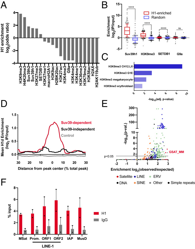
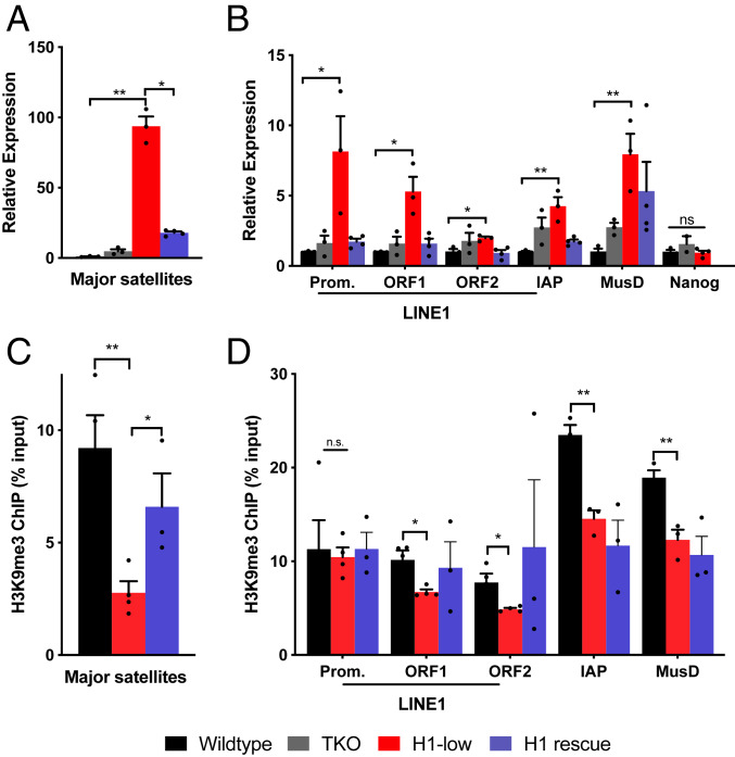
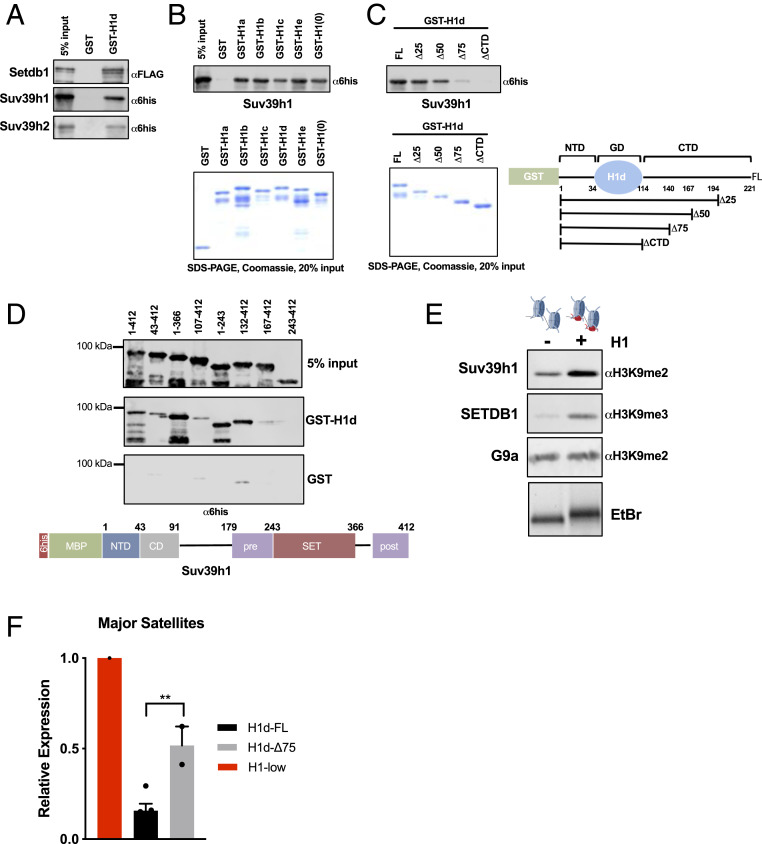
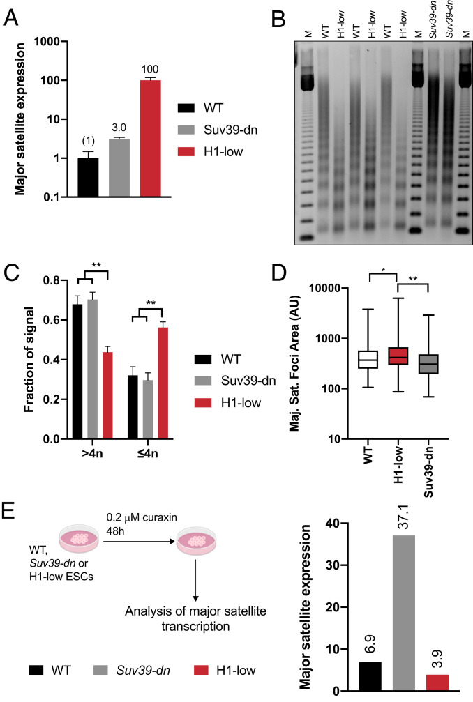
Nearly 50% of mouse and human genomes are composed of repetitive sequences. Transcription of these sequences is tightly controlled during development to prevent genomic instability, inappropriate gene activation and other maladaptive processes. Here, we demonstrate an integral role for H1 linker histones in silencing repetitive elements in mouse embryonic stem cells. Strong H1 depletion causes a profound de-repression of several classes of repetitive sequences, including major satellite, LINE-1, and ERV. Activation of repetitive sequence transcription is accompanied by decreased H3K9 trimethylation of repetitive sequence chromatin. H1 linker histones interact directly with Suv39h1, Suv39h2, and SETDB1, the histone methyltransferases responsible for H3K9 trimethylation of chromatin within these regions, and stimulate their activity toward chromatin in vitro. However, we also implicate chromatin compaction mediated by H1 as an additional, dominant repressive mechanism for silencing of repetitive major satellite sequences. Our findings elucidate two distinct, H1-mediated pathways for silencing heterochromatin.

Keywords: chromatin; epigenetics; linker histones; repetitive elements.


Conflict of interest statement

The authors declare no competing interest.

Figures

Fig. 1.
Fig. 1.
H1 is enriched within constitutive heterochromatin silenced by Suv39h1/2. (A) Overlap of H1d and histone posttranslational modifications or chromatin proteins in mESCs. ChIP-seq data for a number of histone PTMs and chromatin factors in mESCs was analyzed by ISOR to detect enriched regions. The overlap of H1d-enriched domains with each factor or modification is expressed as a log2(odds ratio). (B) The enrichment of Suv39h1, H3K9me3, SETDB1, and G9a within H1-enriched domains was compared to matched control genomic regions of equal size and number. Enrichment is expressed as log2(IP reads/input reads). (C) Significantly enriched histone posttranslational modifications in H1-enriched domains. H1-enriched ISOR regions (P < 0.01; ES >0.5) were analyzed by Enrichr (31) to detect significant overlaps with histone posttranslational modification ChIP-seq datasets. (D) H1 enrichment in Suv39-dependent, Suv39-independent, and control domains. Suv39-dependent and -independent H3K9me3 domains were determined (SI Appendix, Fig. S1 A and B), and the enrichment of H1d within these domains was analyzed using HOMER. Due to variable domain sizes, the x-axis is displayed as a percent of total peak length from the center. (E) Overlap of H1-enriched domains and repetitive elements genome-wide. H1 ISOR domains were analyzed by HOMER, and enrichment is expressed as log2(observed/expected) overlap. (F) Enrichment of H1a at repetitive elements. ChIP-qPCR was performed on fixed chromatin from ES cells using H1a and control IgG antisera. Enrichment is expressed a percentage of input DNA fragment recovered. ns, not statistically significant; *P ≤ 0.05; **P < 0.01; ***P < 0.001; ****P < 0.0001. The Wilcoxon signed-rank test was used to test for significance in B.
Fig. 2.
Fig. 2.
H1 is required for transcriptional repression of several classes repetitive elements. (A and B) Transcript levels of the indicated repetitive sequences were measured by RT-qPCR in WT, TKO, H1-low, and H1 rescue lines stably expressing H1d. Values were normalized to Gapdh mRNA using the ΔΔCt method. Prom., promoter. Error bars represent SEM of three or four independently derived clones per condition. (C and D) H3K9me3 ChIP-qPCR in WT, H1-low, and H1 rescue lines. Cross-linked chromatin was immunoprecipitated with an H3K9me3 antibody, and recovered DNA was quantified by PCR using primers for the indicated repetitive sequences. Data are shown as a percentage of input DNA fragments. Error bars indicate SEM of three or four independent clones per condition. ns, not statistically significant; *P ≤ 0.05; **P < 0.01; ***P < 0.001; ****P < 0.0001.
Fig. 3.
Fig. 3.
Linker histones interact with Suv39h1/h2 and SETDB1 and promote H3K9 methylation in vitro. (A) Interaction studies between H1 and H3K9 methyltransferases. Linker histone GST fusion proteins were purified, bound to glutathione Sepharose beads, and incubated with the indicated recombinant proteins tagged with either hexahistidine-maltose binding protein (6his-MBP) or Flag peptides. Bound proteins and 5% input control samples were analyzed by sodium dodecyl sulfate/polyacrylamide gel electrophoresis (SDS/PAGE) followed by immunoblotting for the indicated peptides. (B and C) GST pulldown assays as in A with the indicated proteins. Coomassie blue-stained SDS/PAGE gel of purified proteins showing equal loading (Lower). (C, Right) Schematic representation of GST-H1d polypeptides used in interaction assays. Numbers indicate amino acid residues. (D) GST pulldown assays as in A with the indicated polypeptide fragments of Suv39h1. After incubation with GST or GST-H1d, pulldown products and 5% input controls were analyzed with a hexahistidine(6his)-specific antibody. A schematic illustration of Suv39h1 domain structure is shown below. (E) In vitro HMT assays with reconstituted chromatin in the presence or absence of H1. Chromatin was reconstituted in vitro using a DNA template bearing two repeats of the synthetic “601” nucleosome positioning sequence and recombinant histone octamers in either the presence or absence of H1. H1 incorporation was verified by nondenaturing agarose gel electrophoresis followed by ethidium bromide (EtBr) staining, demonstrating slower migration of H1-containing dinucleosomes (Lower). Dinucleosomes were incubated with the indicated enzymes (Suv39h1, 100 nM; SETDB1, 50 nM; G9a, 20 nM) under conditions described in Materials and Methods. Enzymatic activity was detected by immunoblotting for the indicated histone modifications. (F) RT-qPCR of major satellite transcripts. Expression of major satellites was quantified in H1-low lines in which either full-length H1d (H1d-FL) or H1d lacking 75% of the CTD (H1d-Δ75) bearing an N-terminal 3xFlag tag were reintroduced via stable transfection. Expression (relative to the parental line) was calculated using the ΔΔCt method normalized to Gapdh mRNA. n = 2 or 3 independent clones per condition.
Fig. 4.
Fig. 4.
H1-mediated chromatin compaction is the dominant mechanism of major satellite repression. (A) Expression of major satellite transcripts in WT, Suv39-dn, and H1-low ES cells. Major satellite transcripts in WT, Suv39-dn, and H1-low ES cells were measured by RT-qPCR and normalized to Gapdh mRNA using the ΔΔCt method. Numbers above indicate expression relative to WT. Data are from three technical replicates of the indicated cell line. Similar results were obtained with several lines of the same genotype (see Fig. 2A). (B) Nucleosome spacing in WT, Suv39-dn, and H1-low ES cells. Nuclei were subjected to limited digestion with micrococcal nuclease and DNA was purified and analyzed by nondenaturing agarose gel electrophoresis, followed by ethidium bromide staining. (C) Analysis of chromatin condensation in WT, Suv39-dn, and H1-low ES cells. The fraction of signal arising from DNA fragments in B corresponding to tetranucleosomes and below (≤4n) and pentanucleosomes and above (>4n) was quantified and normalized to the total signal in each lane using ImageJ. n = 2 or 3 clones per genotype; error bars represent SD. (D) Area of major satellite foci in WT, Suv39-dn, and H1-low ESCs. At 24 h after transfection with TALEN specific to major satellite DNA fused to mClover, cells were sorted, applied to coverslips, and fixed. The area of major satellite foci was determined using ImageJ. WT, n = 180; Suv39-dn, n = 191; H1-low, n = 279. *P ≤ 0.05; **P < 0.01. Unpaired t test with Welch’s correction was used to test statistical significance. (E) Expression of major satellites in dimethyl sulfoxide (DMSO)- and curaxin-treated cells. Major satellite transcripts in WT, Suv39-dn, and H1-low ES cells treated with 0.2 µM curaxin or DMSO for 48 h were measured by RT-qPCR as in A. Data are shown as relative to DMSO control at 24 h.

Comment in

  • Silencing the genome with linker histones.
    Hansen JC. Hansen JC. Proc Natl Acad Sci U S A. 2020 Jul 7;117(27):15388-15390. doi: 10.1073/pnas.2009513117. Epub 2020 Jun 19. Proc Natl Acad Sci U S A. 2020. PMID: 32561644 Free PMC article. No abstract available.

References

    1. Misteli T., Gunjan A., Hock R., Bustin M., Brown D. T., Dynamic binding of histone H1 to chromatin in living cells. Nature 408, 877–881 (2000). - PubMed
    1. Bednar J. et al. ., Structure and dynamics of a 197 bp nucleosome in complex with linker histone H1. Mol. Cell 66, 384–397.e8 (2017). - PMC - PubMed
    1. Woodcock C. L., Skoultchi A. I., Fan Y., Role of linker histone in chromatin structure and function: H1 stoichiometry and nucleosome repeat length. Chromosome Res. 14, 17–25 (2006). - PubMed
    1. Kouzarides T., Chromatin modifications and their function. Cell 128, 693–705 (2007). - PubMed
    1. Happel N., Doenecke D., Histone H1 and its isoforms: Contribution to chromatin structure and function. Gene 431, 1–12 (2009). - PubMed

Publication types

LinkOut - more resources