. 2020 Jul 21;88(8):e00018-20.
doi: 10.1128/IAI.00018-20. Print 2020 Jul 21.

mcr-1 Gene Expression Modulates the Inflammatory Response of Human Macrophages to Escherichia coli

Affiliations

mcr-1 Gene Expression Modulates the Inflammatory Response of Human Macrophages to Escherichia coli

Giorgio Mattiuz et al. Infect Immun. .

Abstract

MCR-1 is a plasmid-encoded phosphoethanolamine transferase able to modify the lipid A structure. It confers resistance to colistin and was isolated from human, animal, and environmental strains of Enterobacteriaceae, raising serious global health concerns. In this paper, we used recombinant mcr-1-expressing Escherichia coli to study the impact of MCR-1 products on E. coli-induced activation of inflammatory pathways in activated THP-1 cells, which was used as a model of human macrophages. We found that infection with recombinant mcr-1-expressing E. coli significantly modulated p38-MAPK and Jun N-terminal protein kinase (JNK) activation and pNF-κB nuclear translocation as well as the expression of genes for the relevant proinflammatory cytokines tumor necrosis factor alpha (TNF-α), interleukin-12 (IL-12), and IL-1β compared with mcr-1-negative strains. Caspase-1 activity and IL-1β secretion were significantly less activated by mcr-1-positive E. coli strains than the mcr-1-negative parental strain. Similar results were obtained with clinical isolates of mcr-1-positive E. coli, suggesting that, in addition to colistin resistance, the expression of mcr-1 allows the escape of early host innate defenses and may promote bacterial survival.

Keywords: MCR-1; cytokines; inflammation; macrophages.


Figures

FIG 1
FIG 1
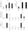
Expression of proinflammatory cytokine genes by PMA/THP-1 cells. PMA/THP-1 cells were cultured in the presence or absence of MCRPEC, MCRNEC, or LPS as a positive control. Results represent the fold change mRNA compared with unstimulated cultures (mean ± SE of triplicate cultures). Data from three representative experiments are shown. Statistical analysis was performed with the Student’s t test. A P value of <0.05 was considered statistically significant.
FIG 2
FIG 2
Production of TNF-α, IL-1β, and IL-10 by PMA/THP-1 cells. PMA/THP-1 cells were cultured in the presence of live bacteria (50 MOI/cell) for 3 hours. Supernatants were collected after 16 hours and analyzed by the immunoplex assay. ANOVA one-way analysis revealed significant differences among cultures with the different strains. The Student’s t test was used to compare cytokine production in cultures with MCRPEC and MCRNEC. A P value of ≤0.05 was considered statistically significant.
FIG 3
FIG 3
NF-κB, p38-MAPK, and SAPK/JNK phosphorylation by PMA/THP-1 cells. PMA/THP-1 cells were cultured for 20 minutes in the absence (US) or presence of MCRPEC, MCRNEC, or LPS as a positive control. Cells were analyzed by Western blot analysis. Data from one representative experiment out of three performed are shown. The bar graph shows the results of densitometric analysis of three different experiments. Data are expressed as mean fold increase ± SE of stimulated cultures over the unstimulated control. Statistical analysis was performed by Student’s t test. A P value of ≤0.05 was considered statistically significant.
FIG 4
FIG 4
Caspase-1 activation and LDH release of PMA/THP-1 cells. (a) PMA/THP-1 cells were cultured in the presence of MCRPEC, MCRNEC, or LPS as a positive control for 6 hours. Caspase-1 activity is expressed as luminescence units (RLUs), as revealed by spectrophotometric analysis. (b) LDH activity in culture supernatants of PMA/THP-1 cells incubated with MCRPEC, MCRNEC, or LPS; data represent mean ± SE of triplicate cultures. (c and d) Caspase-1 activity and LDH production analysis in PMA/THP-1 cells cultured with isolated clinical samples. ANOVA one-way analysis revealed significant differences among cultures. Statistical analysis was performed by Student’s t test. A P value of ≤0.05 was considered significant.

References

    1. Jeannot K, Bolard A, Plésiat P. 2017. Resistance to polymyxins in Gram-negative organisms. Int J Antimicrob Agents 49:526–535. doi:10.1016/j.ijantimicag.2016.11.029. - DOI - PubMed
    1. Trent MS, Stead CM, Tran AX, Hankins JV. 2006. Diversity of endotoxin and its impact on pathogenesis. J Endotoxin Res 12:205–223. doi:10.1179/096805106X118825. - DOI - PubMed
    1. Steimle A, Autenrieth IB, Frick J-S. 2016. Structure and function: lipid A modifications in commensals and pathogens. Int J Med Microbiol 306:290–301. doi:10.1016/j.ijmm.2016.03.001. - DOI - PubMed
    1. Cannatelli A, Giani T, Aiezza N, Di Pilato V, Principe L, Luzzaro F, Galeotti CL, Rossolini GM. 2017. An allelic variant of the PmrB sensor kinase responsible for colistin resistance in an Escherichia coli strain of clinical origin. Sci Rep 7:5071. doi:10.1038/s41598-017-05167-6. - DOI - PMC - PubMed
    1. Bourrel AS, Poirel L, Royer G, Darty M, Vuillemin X, Kieffer N, Clermont O, Denamur E, Nordmann P, Decousser J-W; IAME Resistance Group. 2019. Colistin resistance in Parisian inpatient faecal Escherichia coli as the result of two distinct evolutionary pathways. J Antimicrob Chemother 74:1521–1530. doi:10.1093/jac/dkz090. - DOI - PubMed

Publication types

MeSH terms