. 2020 Jul 1;12(550):eabc3539.
doi: 10.1126/scitranslmed.abc3539. Epub 2020 Jun 8.

Antibody signature induced by SARS-CoV-2 spike protein immunogens in rabbits

Affiliations

Antibody signature induced by SARS-CoV-2 spike protein immunogens in rabbits

Supriya Ravichandran et al. Sci Transl Med. .

Abstract

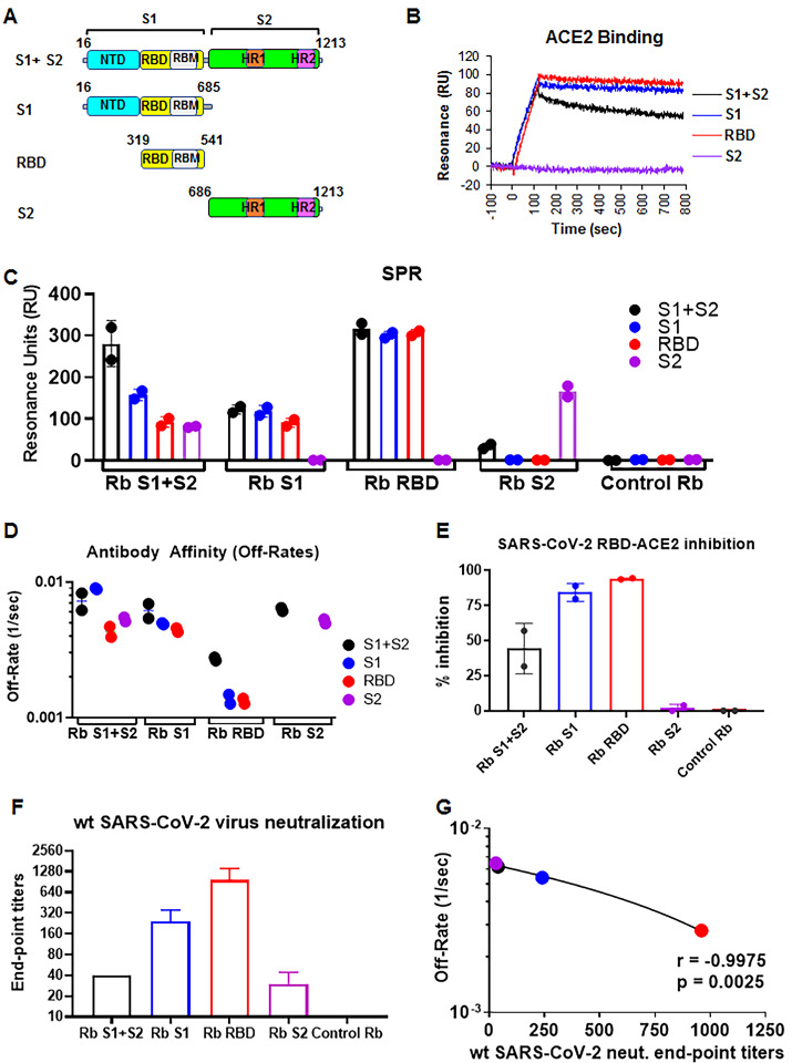
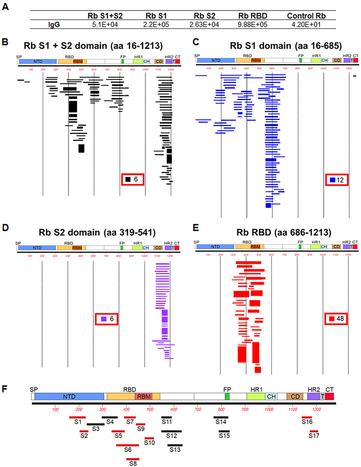
Multiple vaccine candidates against SARS-CoV-2 based on viral spike protein are under development. However, there is limited information on the quality of antibody responses generated with these vaccine modalities. To better understand antibody responses induced by spike protein-based vaccines, we performed a qualitative study by immunizing rabbits with various SARS-CoV-2 spike protein antigens: S ectodomain (S1+S2; amino acids 16 to 1213), which lacks the cytoplasmic and transmembrane domains (CT-TM), the S1 domain (amino acids 16 to 685), the receptor binding domain (RBD) (amino acids 319 to 541), and the S2 domain (amino acids 686 to 1213, lacking the RBD, as control). Resulting antibody quality and function were analyzed by enzyme-linked immunosorbent assay (ELISA), RBD competition assay, surface plasmon resonance (SPR) against different spike proteins in native conformation, and neutralization assays. All three antigens (S1+S2 ectodomain, S1 domain, and RBD), but not S2, generated strong neutralizing antibodies against SARS-CoV-2. Vaccination-induced antibody repertoire was analyzed by SARS-CoV-2 spike genome fragment phage display libraries (SARS-CoV-2 GFPDL), which identified immunodominant epitopes in the S1, S1-RBD, and S2 domains. Furthermore, these analyses demonstrated that the RBD immunogen elicited a higher antibody titer with five-fold higher affinity antibodies to native spike antigens compared with other spike antigens, and antibody affinity correlated strongly with neutralization titers. These findings may help guide rational vaccine design and facilitate development and evaluation of effective therapeutics and vaccines against COVID-19 disease.


Figures

Fig. 1
Fig. 1. SARS-CoV-2 spike binding and SARS-CoV-2 neutralization by serum antibodies generated following rabbit immunization with spike antigens.
A) Schematic representation of the SARS-CoV-2 spike protein and subdomains. Spike S1+S2 ectodomain (aa 16-1213) lacks the cytoplasmic and transmembrane domains (CT-TM), S1 domain (aa 16-685), RBD domain (aa 319-541), and S2 domain (aa 686-1213), all containing 6x His tag at C terminus, were commercially produced in either HEK 293 mammalian cells (S1 and RBD) or insect cells (S1+S2 ectodomain and S2 domain). The receptor-binding motif (RBM) encompasses residues 437-508. (B) Binding of purified proteins to human ACE2 (hACE2) proteins in SPR. Sensorgrams represent binding of purified spike proteins on low-density His-captured chips to 5 μg/mL human ACE2 protein. (C) SPR binding of antibodies from two rabbits each immunized twice with SARS-CoV-2 antigens to spike protein and domains from SARS-CoV-2 (S1+S2, black; S1, blue; RBD, red; and S2, purple). Total antibody binding is represented in maximum resonance units (RU) in this figure for 10-fold serum dilution. All SPR experiments were performed twice and the researchers performing the assay were blinded to sample identity. The variations for duplicate runs of SPR was <5%. The data shown are average values of two experimental runs. (D) Antibody off-rate constants were determined directly from the serum sample interaction with SARS-CoV-2 spike ectodomain (S1+S2), S1, S2, and RBD using SPR in the dissociation phase only for the sensorgrams with Max RU in the range of 20–100 RU. (E) RBD-hACE2 competition assay. Percent inhibition of hACE2 binding to RBD in presence of 1:50 dilutions of post-second vaccination rabbit serum was measured by SPR. (F) End-point virus neutralization titers for one rabbit from each group using wild type SARS-CoV-2 virus in a classical BSL3 neutralization assay based on CPE (Cytopathic effect) was performed as described in Materials and Methods. (G) Anti-spike ectodomain (S1+S2) binding antibody affinity as measured by antibody dissociation rates (off-rates) of post-vaccinated rabbit polyclonal antibodies correlated with the wt SARS-CoV-2 virus end-point neutralization titers (r = -0.9975, p <0.005). Pearson two-tailed correlations are reported for the calculation of correlations between anti-S1+S2 antibody affinity and end-point titers for one rabbit per immunogen. The color scheme in panel G is the same as in panel D/F.
Fig. 2
Fig. 2. Antibody epitope repertoires generated by different SARS-CoV-2 spike antigens.
(A) Number of IgG-bound SARS-CoV-2 GFPDL phage clones using the post-second vaccination rabbit polyclonal sera. (B-E) Graphical distribution of representative clones with a frequency of ≥2, obtained after affinity selection, and their alignment to the spike protein of SARS-CoV-2 are shown for the four vaccine groups: S1+S2 ectodomain (B), S1 (C), S2 domain (D) and S1-receptor binding domain (RBD) (E). The thickness of each bar represents the frequency of repetitively isolated phage, with the scale shown enclosed in a red box in the respective alignments in each panel. (F) Elucidation of the antibody epitope profile in SARS-CoV-2 spike following rabbit vaccination. Antigenic sites within the SARS-CoV-2 spike protein recognized by serum antibodies following rabbit vaccination (based on data presented in Fig. 1B-E). The amino acid designation is based on the SARS-CoV-2 spike protein sequence (fig. S4). The antigenic regions/sites are depicted below the spike schematic and are color coded. Epitopes of each protein are numbered in a sequential fashion indicated in black. The antigenic epitopes are color coded unique to this study (red bars) or if they were predicted by algorithms (black bars) previously by Grifoni et al. (19). Sequence residues for each antigenic site and their sequence conservation with other human coronaviruses is shown in table S1. The GFPDL affinity selection data was performed twice. Similar numbers of phage clones and epitope repertoire were observed in both phage display analyses.

References

    1. John Hopkins University & Medicine Center for Systems Science and Engineering (CSSE), (2020), vol. 2020.
    1. Kuba K., Imai Y., Rao S., Gao H., Guo F., Guan B., Huan Y., Yang P., Zhang Y., Deng W., Bao L., Zhang B., Liu G., Wang Z., Chappell M., Liu Y., Zheng D., Leibbrandt A., Wada T., Slutsky A. S., Liu D., Qin C., Jiang C., Penninger J. M., A crucial role of angiotensin converting enzyme 2 (ACE2) in SARS coronavirus-induced lung injury. Nat. Med. 11, 875–879 (2005). 10.1038/nm1267 - DOI - PMC - PubMed
    1. Wrapp D., Wang N., Corbett K. S., Goldsmith J. A., Hsieh C. L., Abiona O., Graham B. S., McLellan J. S., Cryo-EM structure of the 2019-nCoV spike in the prefusion conformation. Science 367, 1260–1263 (2020). 10.1126/science.abb2507 - DOI - PMC - PubMed
    1. Yan R., Zhang Y., Li Y., Xia L., Guo Y., Zhou Q., Structural basis for the recognition of SARS-CoV-2 by full-length human ACE2. Science 367, 1444–1448 (2020). 10.1126/science.abb2762 - DOI - PMC - PubMed
    1. Walls A. C., Park Y. J., Tortorici M. A., Wall A., McGuire A. T., Veesler D., Structure, Function, and Antigenicity of the SARS-CoV-2 Spike Glycoprotein. Cell 181, 281–292.e6 (2020). 10.1016/j.cell.2020.02.058 - DOI - PMC - PubMed

Publication types