. 2020 Jun 8;10(1):9166.
doi: 10.1038/s41598-020-65983-1.

Abnormal Papillary Muscle Signal on Cine MRI As a Typical Feature of Mitral Valve Prolapse

Affiliations

Abnormal Papillary Muscle Signal on Cine MRI As a Typical Feature of Mitral Valve Prolapse

Alessandra Scatteia et al. Sci Rep. .

Abstract

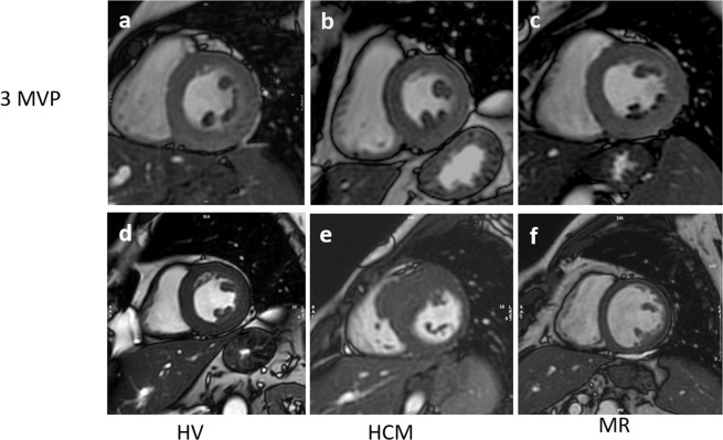
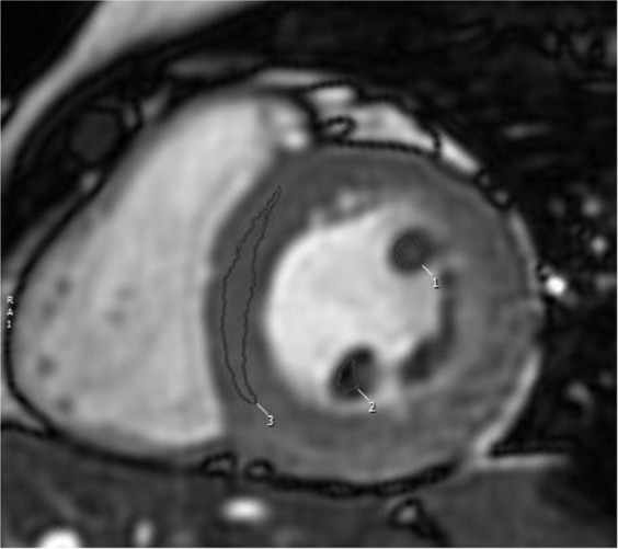
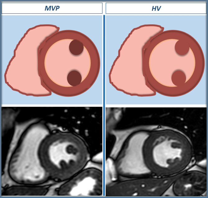
Background: Mitral valve prolapse (MVP) is characterized by an abnormal movement of the valvular apparatus which may affect the papillary muscles (PMs) function and structure. Aim of the study was to investigate abnormal PM signal in MVP by using cardiac magnetic resonance imaging (MRI).

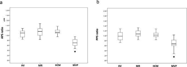
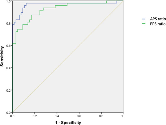
Methods and results: We enrolled 47 consecutive patients with MVP evaluated by cardiac MRI. Additional groups included healthy volunteers, patients with moderate-to-severe mitral regurgitation (not caused by MVP) and patients with hypertrophic cardiomyopathy. Visual assessment of the PM signals was carried out and the signal intensity (SI) of both the antero-lateral and postero-medial PMs was normalized by that of the left ventricular (LV) parietal myocardium. Our results show that in the MVP group only, the PM signal intensity was significantly lower compared to the one of the LV parietal myocardium. This sign did not correlate with either LV late gadolinium enhancement or positive anamnesis for significant arrhythmias.

Conclusions: In MVP patients only, PM signal is significantly reduced compared to LV parietal myocardium ("darker appearance"). The described findings are not clearly related to evidence of myocardial fibrosis, as assessed by MRI, and to previous occurrence of complex ventricular arrhythmias.


Conflict of interest statement

Dr. Scatteia and Dr. Guarini are partially funded from the “Centro Studi della Società di Cardiologia Ospedaliera Accreditata (SICOA)”. The remaining authors declare no competing interests.

Figures

Figure 1
Figure 1
Examples of end-systolic short-axis cine images with clearly lower papillary muscle signal intensity as compared to parietal left ventricular myocardium in MVP patients (ac), but not in HV(d), HCM (e) and MR (f).
Figure 2
Figure 2
Manually drawing of appropriate region of interest (ROI) on papillary muscle and parietal left ventricle for signal intensity calculation. Post processing with CVi42, Circle Cardiovascular Imaging Inc., Calgary, Canada.
Figure 3
Figure 3
Values of anterior papillary signal (APS) ratio (a) and posterior papillary signal (PPS) ratio (b) as measured in the different study groups. HCM: Hypertrophic cardiomyopathy group; HV: Healthy volunteers group; MR: Mitral regurgitation group; MVP: Mitral valve prolapse group. Median and interquartile range are indicated. *p value <0,001 between MVP patients and all the other groups.
Figure 4
Figure 4
ROC curve analysis for the diagnostic performance of anterior papillary signal (APS) ratio and posterior papillary signal (PPS) ratio in identifying patients with mitral valve prolapse. Area under the curve is 0,96 for APS ratio and 0,92 for PPS ratio.
Figure 5
Figure 5
Schematic representation of papillary muscles signal intensity, with correspondent end-systolic short-axis cine images in a MVP case and a HV case.

References

    1. Delling FN, et al. Evolution of Mitral Valve Prolapse. Circulation. 2016;133:1688–1695. doi: 10.1161/CIRCULATIONAHA.115.020621. - DOI - PMC - PubMed
    1. Parwani P, Avierinos J-F, Levine RA, Delling FN. Mitral Valve Prolapse: Multimodality Imaging and Genetic Insights. Prog. Cardiovasc. Dis. 2017;60:361–369. doi: 10.1016/j.pcad.2017.10.007. - DOI - PMC - PubMed
    1. Sanfilippo AJ, et al. Papillary Muscle Traction in Mitral Valve Prolapse: Quantitation by Two-Dimensional Echocardiography. J. Am. Coll Cardiol. 1992;19:564–71. doi: 10.1016/S0735-1097(10)80274-8. - DOI - PubMed
    1. Winkle RA, et al. Arrhythmias in patients with mitral valve prolapse. Circulation. 1975;52:73–81. doi: 10.1161/01.CIR.52.1.73. - DOI - PubMed
    1. Basso C, et al. Arrhythmic Mitral Valve Prolapse and Sudden Cardiac Death. Circulation. 2015;132:556–66. doi: 10.1161/CIRCULATIONAHA.115.016291. - DOI - PubMed

MeSH terms