. 2020 Jun 1:6:13.
doi: 10.1186/s42234-020-00048-2. eCollection 2020.

Ultrasound-driven piezoelectric current activates spinal cord neurocircuits and restores locomotion in rats with spinal cord injury

Affiliations

Ultrasound-driven piezoelectric current activates spinal cord neurocircuits and restores locomotion in rats with spinal cord injury

Shuai Li et al. Bioelectron Med. .

Abstract

Background: Neuromodulation via electrical stimulation (ES) is a common technique to treat numerous brain and spinal cord related neurological conditions. In the present study, we examined the efficacy of piezoelectric stimulation (pES) by a custom miniature piezostimulator to activate the spinal cord neurocircuit in comparison with conventional epidural ES in rats.

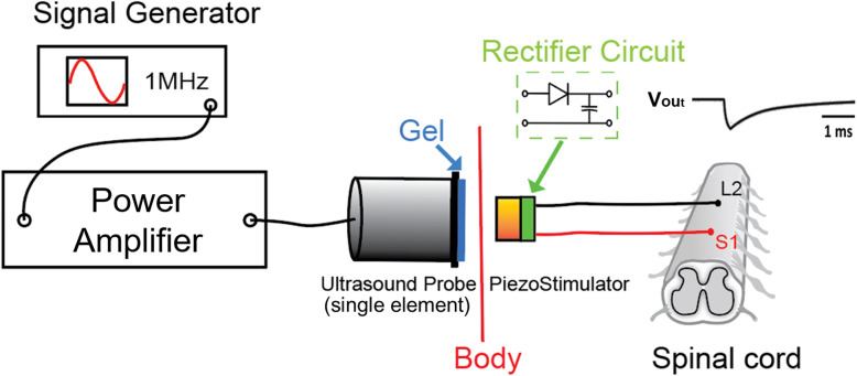
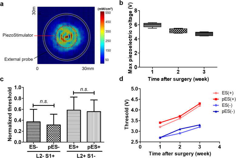
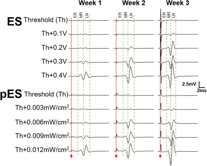
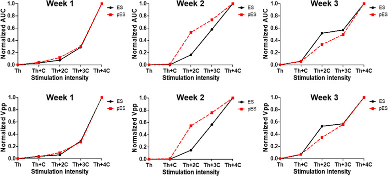
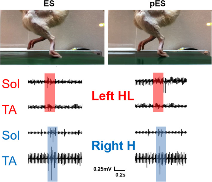
Methods: Stimulation electrodes were implanted on L2 and S1 spinal cord and were connected to a head-plug for ES, and a piezostimulator for pES. EMG electrodes were implanted into hindlimb muscles. To generate piezoelectric current, an ultrasound beam was delivered by an external ultrasound probe. Motor evoked potentials (MEPs) were recorded during the piezoelectric stimulation and compared with the signals generated by the ES.

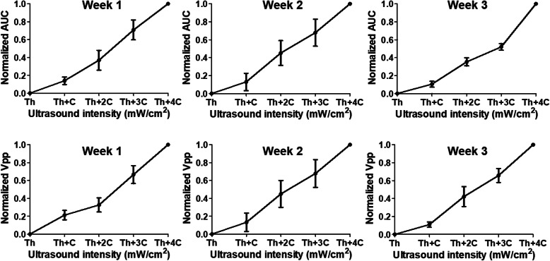
Results: Our results suggest that ultrasound intensity as low as 0.1 mW/cm2 could induce MEPs in the hindlimbs. No significant difference was found either in MEPs or in muscle recruitments for ES and pES. Similar to ES, pES induced by 22.5 mW/cm2 ultrasound restored locomotion in paralyzed rats with complete thoracic cord injury. Locomotion EMG signals indicated that pES works same as ES.

Conclusion: We propose piezoelectric stimulation as a new avenue of neuromodulation with features overtaking conventional electrical stimulation to serve future bioelectronic medicine. Video abstract.

Keywords: Epidural; Neuromodulation, piezoelectric; Neurostimulation; Spinal cord injury; Ultrasound.


Conflict of interest statement

Competing interestsThe authors S.L., M.A., X.Y.W and Y.P.Z are inventors of related patents owned by the Hong Kong Polytechnic University. The remaining authors declare no competing financial interests.

Figures

Fig. 1
Fig. 1
Schematic diagram of the piezoelectric stimulation (pES) experiment setup. The setup includes an external ultrasound energy transmitting system and implanted receiving and piezoelectric current generation module. A signal generator produces 200 μs bursts (1 MHz carrier frequency) of sinusoidal signal and feeds it into a power amplifier to drive an ultrasound probe to produce acoustic energy. With the aid of ultrasound gels, the acoustic energy was then transferred through the skin and was received by the piezostimulator implant. The implant contained a rectifier and filter circuit to convert the sinusoidal piezoelectric signal into a monophasic stimulation pulse for pES. This pES pulse was then delivered to the lumbosacral spinal cord (L2 and S1 levels) via Teflon-coated stimulation wires to activate the neural circuits
Fig. 2
Fig. 2
a Under PRF of 1 kHz, acoustic intensity-distributing map of the external ultrasound probe measured at a distance of 4 mm from its surface. The black circle indicates the size of ultrasound probe, and the red dotted circle indicates the size of the implanted piezoelectric stimulator. Acoustic intensities ISPTA (spatial-peak temporal-average) was 561.9 mW/cm2 and ISPPA (spatial-peak pulse-average) was 3.9 W/cm2 (Mechanical Index, MI was 0.26). b Maximum harvested piezoelectric pulse voltage from all healthy rats (n = 5). Maximum pES voltage was found to be stable for the first 3 weeks after implantation. Scar tissue formation and change in acoustic impedance might be the cause of the decreased piezoelectric voltage. c Comparisons between conventional electrical stimulation (ES) and pES threshold voltages to activate L2 and S1 spinal cord segments (both anodic: current from L2 to S1, and cathodic: current from S1 to L2). No significant difference was found in the threshold voltages between ES and pES for either anodic or cathodic stimulations. d Trends of ES and pES voltage thresholds over 3 weeks post-implantation in one intact rat. Both stimulation thresholds tend to increase over time, but clearly followed each other (ES vs. pES)
Fig. 3
Fig. 3
Motor evoked potentials (MEPs) of the tibialis anterior (TA) muscle activated by ES and pES stimulation pulses in the first 3 weeks after surgery. For different weeks, the stimulation threshold was different for both ES and pES. In each stimulation type (ES and pES), a constant input intensity (voltage or acoustic strength) was used to find if there was any difference of their MEPs. For ES, 0.1 V was the constant step of intensity, and ISPTA of 0.003 mW/cm2 was the constant step of intensity for pES. Stimulation onset is indicated by a red line. Early (ER), middle (MR) and late (LR) responses are also marked with dotted lines
Fig. 4
Fig. 4
Normalized area-under-the-curve (AUC) and peak-to-peak voltage (Vpp) were measured to enable a clear comparison between ES and pES MEPs. Week 1 and week 3 AUC and Vpp show similar recruitment curves of both ES and pES, while week 2 spinal cord seems to be more sensitive to the pES. C equals to 0.1 V for ES or 0.003 mW/cm2 for pES
Fig. 5
Fig. 5
Normalized AUC and Vpp of MEPs collected from all healthy rats (n = 5) for 3 weeks post-surgery. Recruitment curves of AUC and Vpp with increase of acoustic intensity indicate increase in movement strengths via pES current. C equals 0.003 mW/cm2 for pES
Fig. 6
Fig. 6
Demonstration of a paralyzed rat for successful movement restoration of hindlimbs on a moving treadmill belt during conventional ES and our pES pulses at 40 Hz. EMG signals were recorded from the soleus and TA muscles for both ES and pES, and are shown within one complete gait cycle

Comment in

References

    1. Alam M, Li S, Ahmed RU, Yam YM, Thakur S, Wang XY, Tang D, Ng S, Zheng YP. Development of a battery-free ultrasonically powered functional electrical stimulator for movement restoration after paralyzing spinal cord injury. J Neuroeng Rehabil. 2019. 10.1186/s12984-019-0501-4. - PMC - PubMed
    1. Amar AB, Kouki AB, Cao H. Power approaches for implantable medical devices. Sensors. 2015. 10.3390/s151128889. - PMC - PubMed
    1. Edgerton VR, Courtine G, Gerasimenko YP, Lavrov I, Ichiyama RM, Fong AJ, Cai LL, Otoshi CK, Tillakaratne NJ, Burdick JW. Training locomotor networks. Brain Res Rev. 2008. 10.1016/j.brainresrev.2007.09.002. - PMC - PubMed
    1. Feigin VL, Vos T, Nichols E, Owolabi MO, Carroll WM, Dichgans M, et al. The global burden of neurological disorders: translating evidence into policy. Lancet Neurol. 2020;19:255–65. https://www.ncbi.nlm.nih.gov/pubmed/31813850. - PMC - PubMed
    1. Food and Drug Administration . Marketing Clearance of Diagnostic Ultrasound Systems and Transducers. 2019.

LinkOut - more resources