. 2020 Aug 1;29(15):953-966.
doi: 10.1089/scd.2020.0075. Epub 2020 Jul 2.

Genetic Mouse Models and Induced Pluripotent Stem Cells for Studying Tracheal-Esophageal Separation and Esophageal Development

Affiliations

Genetic Mouse Models and Induced Pluripotent Stem Cells for Studying Tracheal-Esophageal Separation and Esophageal Development

Suleen Raad et al. Stem Cells Dev. .

Abstract

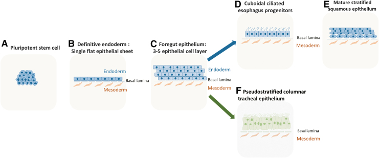
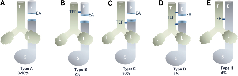
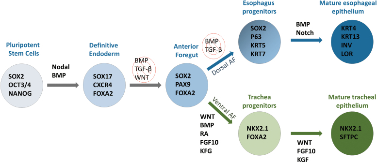
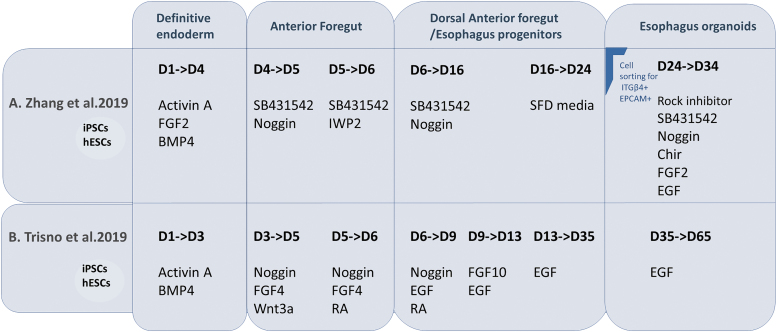
Esophagus and trachea arise from a common origin, the anterior foregut tube. The compartmentalization process of the foregut into the esophagus and trachea is still poorly understood. Esophageal atresia/tracheoesophageal fistula (EA/TEF) is one of the most common gastrointestinal congenital defects with an incidence rate of 1 in 2,500 births. EA/TEF is linked to the disruption of the compartmentalization process of the foregut tube. In EA/TEF patients, other organ anomalies and disorders have also been reported. Over the last two decades, animal models have shown the involvement of multiple signaling pathways and transcription factors in the development of the esophagus and trachea. Use of induced pluripotent stem cells (iPSCs) to understand organogenesis has been a valuable tool for mimicking gastrointestinal and respiratory organs. This review focuses on the signaling mechanisms involved in esophageal development and the use of iPSCs to model and understand it.

Keywords: esophageal atresia/tracheoesophageal fistula; esophageal organoids; induced pluripotent stem cells.


Conflict of interest statement

No competing financial interests exist.

Figures

FIG. 1.
FIG. 1.
Development of esophagus and congenital anomalies: (A) at weeks 4–5 of human embryonic development, the anterior foregut tube is patterned down the sagittal axis to separate dorsally into the esophagus and ventrally into the trachea. (B) The ventral foregut endoderm evaginates and pushes the surrounding mesenchyme to initiate lung bud formation. A unique epithelial population at the D/V midline fuses to form a septum. (C) The separation between ventral and dorsal endoderm starts and progresses in a rostral direction. (D) Intact normal separation of the esophagus in blue and trachea in green. (E) Patterning anomalies lead to the development of congenital anomalies such as EA/TEF. D/V, dorsal-ventral; E, esophagus; EA, esophageal atresia; L, lungs; S, stomach; T, trachea; TEF, tracheoesophageal fistula. Color images are available online.
FIG. 2.
FIG. 2.
Epithelial morphogenesis of the esophagus and trachea: (A) pluripotent stem cells with characteristics of high nucleus-cytoplasm ratio, prominent nucleoli, and formation of compact colonies. (B) The DE is made up of a single flat epithelial sheet with a supporting mesoderm. (C) 3–5 epithelial cell layer makes up the foregut epithelium, which is then patterned along the D/V axis to give rise to (D) esophageal cuboidal ciliated progenitor epithelium. (E) Esophageal progenitors then further mature to become a stratified squamous epithelium with a basal proliferative layer and a suprabasal layer and (F) tracheal pseudostratified columnar tracheal epithelium. DE, definitive endoderm. Color images are available online.
FIG. 3.
FIG. 3.
Types of EA and TEF: (A) type A, a less common subtype of EA/TEF where the esophagus is discontinued without connecting the trachea. (B) Type B, one of the least common subtypes of EA/TEF where a fistula connects the upper part of the esophagus to the trachea and the esophagus ends in a blind pouch. (C) The most common type of EA/TEF occurring in 80% of the cases where the esophagus is discontinued and TEF occurs in the lower section of the esophagus. (D) Type D is the least common subtype of EA/TEF where there is EA with two fistulas between the trachea and the lower and upper esophageal pouches. (E) Type H is one of the less common subtypes of EA/TEF occurring in 4% of the cases where the esophagus connects normally to the stomach with a fistula connecting the esophagus to the stomach. Color images are available online.
FIG. 4.
FIG. 4.
Signaling networks responsible for D/V patterning and subsequent separation of the anterior foregut into either esophageal or respiratory fate: (A) patterning of the anterior foregut tube is regulated by a complex communication network between the mesenchyme (in orange) and the endoderm (in blue or green). Main signaling pathways such as BMP, WNT, FGF, and SHH are key in establishing patterning in the AFE. Any disruption of these signaling pathways may lead to segregation defects. (B) Following patterning, segregation of the foregut tube is established by a specific epithelial population SOX2+NKX2.1+ISL1+orchestrated by mesenchymal cell invasion and localized extracellular matrix degradation allowing the intact separation of the foregut tube into the esophagus on the dorsal side and the trachea on the ventral side. AFE, anterior foregut epithelium; BMP, bone morphogenetic protein; FGF, fibroblast growth factor; SHH, sonic hedgehog. Color images are available online.
FIG. 5.
FIG. 5.
Scheme representing protocols for differentiation of iPSCs/hESCs into mature esophageal and tracheal epithelium: the first critical step is the formation of the DE from pluripotent stem cells expressing SOX2, OCT3/4, and NANOG. DE is generated by activating the Nodal and BMP pathways and the expression of SOX17, CXCR4, and FOXA2. DE cell population is then directed into an AF fate by the inhibition of BMP, TGF-β, and WNT pathways. Trachea and esophagus derive from the anterior side of the foregut. Patterning of the anterior foregut tube along sagittal axis results in the formation of the esophagus on the dorsal side and the trachea on the ventral side. By mimicking the main signaling pathways involved in foregut patterning, AF can generate esophageal progenitors by the dual inhibition of BMP and TGF-β pathways and the tracheal progenitors by activation of WNT, BMP, and RA signaling pathways. Esophageal progenitors can be further matured by activating the BMP and Notch signaling pathways to generate a stratified squamous epithelium expression KRT4, KRT13, INV, and LOR. On the other hand, tracheal progenitors generate mature tracheal epithelium expressing NKX2.1 and SFTC by activating the Wnt pathway. AF, anterior foregut; hESCs, human embryonic stem cells; iPSCs, induced pluripotent stem cells; TGF-β, tumor growth factor-β. Color images are available online.
FIG. 6.
FIG. 6.
Schematic presentation of two published differentiation protocols of iPSCs/hESCs into esophageal organoids: (A) Zhang et al. describe a step-wise protocol where they differentiate iPSCs/hESCs into DE using Activin A, FGF2, and BMP4 to form embryoid bodies that are further induced into anterior foregut by culturing in media supplemented with SB431542, Noggin followed by IWP2 to inhibit the TGF-β, BMP, and Wnt pathways, respectively. After esophageal fate commitment, cells were sorted (ITGβ4+EPCAM+) and resuspended to form esophageal organoids with an organoid culture medium with specific growth factors. (B) Trisno et al., propose a differentiation protocol where iPSCs/hESCs are subjected to Activin A and BMP4 for 3 days to generate DE. Anterior foregut spheroids are then generated by inhibiting the BMP pathways and activating the Wnt pathways for two consecutive days followed by inhibition of BMP and activation of RA for 24 h. AF spheroids were then plated on Matrigel and dorsal AF patterning was achieved by the continued inhibition of BMP and activation of RA, in addition to supplementing with EGF and FGF10. This favored the efficiency of spheroid to organoid outgrowth. Esophageal organoids were maintained for up to 2 months. Activin A, activates Nodal Pathway; BMP4, activates bone morphogenetic proteins pathway; Chir, activates wingless-related integration site pathway; EGF, activates epidermal growth factor pathway; FGF2, FGF4, and FGF10, activate Fibroblast Growth pathway; IWP2, inhibits wingless-related integration site pathway; Noggin, inhibits bone morphogenetic proteins pathway; RA, activates retinoic acid pathway; Rock inhibitor, Y-27632 selectively inhibits p160ROCK to increase survivability; SB431542, inhibits transformation growth factor-β pathway; SFD, serum-free differentiation media. Color images are available online.

Similar articles

Cited by

References

    1. Jacobs IJ, Ku WY and Que J. (2012). Genetic and cellular mechanisms regulating anterior foregut and esophageal development. Dev Biol 369:54–64. - PMC - PubMed
    1. Yu WY, Slack JM and Tosh D. (2005). Conversion of columnar to stratified squamous epithelium in the developing mouse oesophagus. Dev Biol 284:157–170. - PubMed
    1. Perl AK, Wert SE, Nagy A, Lobe CG and Whitsett JA. (2002). Early restriction of peripheral and proximal cell lineages during formation of the lung. Proc Natl Acad Sci U S A 99:10482–10487. - PMC - PubMed
    1. van Lennep M, Singendonk MMJ, Dall'Oglio L, Gottrand F, Krishnan U, Terheggen-Lagro SWJ, Omari TI, Benninga MA and van Wijk MP. (2019). Oesophageal atresia. Nat Rev Dis Primers 5:26. - PubMed
    1. Peters JH and Avisar N. (2010). The molecular pathogenesis of Barrett's esophagus: common signaling pathways in embryogenesis metaplasia and neoplasia. J Gastrointest Surg 14(Suppl. 1):S81–S87. - PubMed

Publication types

LinkOut - more resources