. 2020 Sep 16;75(9):e53-e62.
doi: 10.1093/gerona/glaa121.

In Vivo Quasi-Elastic Light Scattering Eye Scanner Detects Molecular Aging in Humans

Affiliations

In Vivo Quasi-Elastic Light Scattering Eye Scanner Detects Molecular Aging in Humans

Olga Minaeva et al. J Gerontol A Biol Sci Med Sci. .

Abstract

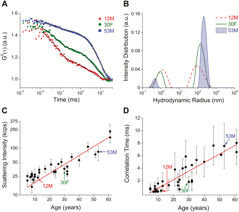
The absence of clinical tools to evaluate individual variation in the pace of aging represents a major impediment to understanding aging and maximizing health throughout life. The human lens is an ideal tissue for quantitative assessment of molecular aging in vivo. Long-lived proteins in lens fiber cells are expressed during fetal life, do not undergo turnover, accumulate molecular alterations throughout life, and are optically accessible in vivo. We used quasi-elastic light scattering (QLS) to measure age-dependent signals in lenses of healthy human subjects. Age-dependent QLS signal changes detected in vivo recapitulated time-dependent changes in hydrodynamic radius, protein polydispersity, and supramolecular order of human lens proteins during long-term incubation (~1 year) and in response to sustained oxidation (~2.5 months) in vitro. Our findings demonstrate that QLS analysis of human lens proteins provides a practical technique for noninvasive assessment of molecular aging in vivo.

Keywords: Molecular aging; Crystallin; Human; Lens; Protein aggregation.


Figures

Figure 1.
Figure 1.
Long-lived proteins in the human body. (A) A small number of proteins are expressed during development and remain extant throughout life. These long-lived proteins include: collagen, the most abundant protein in the body, found in bone, tendon, cartilage, muscle, intervertebral discs, skin, and cornea; enamel and dentin proteins in teeth; nuclear pore proteins (nucleoporins) in neurons in the brain; elastin in heart, lung, and blood vessels; and the crystallins (α, β, γ), cytosolic proteins in the lens of the eye. (B) Diagrammatic representation of the human lens in transverse section (left) and magnified coronal section at the equator (right). (C) Long-lived structural proteins are subject to cumulative molecular damage, posttranslational alterations, and supramolecular reorganization, including pathogenic protein aggregation. Collectively, these molecular pathologies impair organelle, cellular, and tissue homeostasis; degrade structural and physiological functions; and accelerate biological aging.
Figure 2.
Figure 2.
Quasi-elastic light scattering (QLS) instrument and performance characteristics. (A) Optical diagram of QLS instrument. CL, cylindrical lens; BS, beam splitter; GL, GRIN lens. (B) Particle size distributions extracted from the mean autocorrelation functions obtained by QLS analysis of 50-nm radius polystyrene beads in water. Calculated mean hydrodynamic radius (48 nm) closely approximates mean bead radius. (C) Scattering intensity of light increases linearly with concentration of 50-nm radius polystyrene beads suspended in water. Each point represents mean value ± SD. (D) Correlation time increases linearly with polystyrene bead radius. Each point represents mean value ± SD.
Figure 3.
Figure 3.
Molecular aging detected by quasi-elastic light scattering (QLS) in vivo. (A) Representative QLS autocorrelation functions obtained from three healthy human subjects (12-year-old male, 30-year-old female, 53-year-old male). (B) Shift in hydrodynamic radius for the same subjects. (C) Increase in light scattering intensity as a function of age in healthy human subjects (N = 34; 18 males, 16 females). Data collected from nuclear region of the lens. Red line, exponential fit model, R2 = 0.83 (calculated for log-transformed data). (D) Change in correlation time observed with increasing age. Red line, linear fit model, R2 = 0.81.
Figure 4.
Figure 4.
Quasi-elastic light scattering (QLS) detects time-dependent changes in human lens protein during long-term (~1 year) incubation in vitro. (A) Increase in light scattering intensity during long-term (~1 year) incubation of human lens protein in vitro. Red line, exponential fit model (R2 = 0.99). (B) Increase in correlation time during long-term (~1 year) incubation in vitro. Red line, linear fit model (R2 = 0.99). Black arrow, calculated time of inflection. Red arrow, apparent reduction in correlation time. (C) Mean particle size distribution increases with incubation time.
Figure 5.
Figure 5.
Oxidation accelerates time-dependent quasi-elastic light scattering (QLS) changes in human lens protein during long-term incubation in vitro. (A) Change in light scattering intensity in control and oxidized human lens protein samples as a function of incubation time in vitro. (B) Change in correlation time in control and oxidized human lens protein samples as a function of incubation time. Error bars represent SD. Red arrow, apparent reduction in correlation time. (C–E) Protein gel electrophoresis (left panels), gel band densitometry plots (middle panels), and transmission electron microscope (TEM) images (right panels) comparing control and oxidized human lens protein specimens at representative time points during incubation (C, Day 0; D, Day 36; E, Day 77). Gel band densitometry plots are normalized to total signal for each sample. Arrow, high-molecular-weight (HMW) lens protein. Box, lens protein bands corresponding to α-crystallins. Histogram color code: violet = 0–11 kDa; blue = 12–32 kDa; green = 33–89 kDa; orange = 90–255 kDa; red > 255 kDa. Scale bar (TEM) = 100 nm.

References

    1. Herndon LA, Schmeissner PJ, Dudaronek JM, et al. . Stochastic and genetic factors influence tissue-specific decline in ageing C. elegans. Nature. 2002;419:808–814. doi:10.1038/nature01135 - DOI - PubMed
    1. Kirkwood TB, Feder M, Finch CE, et al. . What accounts for the wide variation in life span of genetically identical organisms reared in a constant environment? Mech Ageing Dev. 2005;126:439–443. doi:10.1016/j.mad.2004.09.008 - DOI - PubMed
    1. Hayflick L. Biological aging is no longer an unsolved problem. Ann NY Acad Sci. 2007;1100:1–13. doi:10.1196/annals.1395.001 - DOI - PubMed
    1. Johnson TE. Recent results: biomarkers of aging. Exp Gerontol. 2006;41:1243–1246. doi:10.1016/j.exger.2006.09.006 - DOI - PubMed
    1. Sanderson WC, Scherbov S. Demography. Remeasuring aging. Science. 2010;329:1287–1288. doi:10.1126/science.1193647 - DOI - PubMed

Publication types