. 2020 Jun 9;12(11):10129-10146.
doi: 10.18632/aging.103310. Epub 2020 Jun 9.

Clusterin exerts a cytoprotective and antioxidant effect in human osteoarthritic cartilage

Affiliations

Clusterin exerts a cytoprotective and antioxidant effect in human osteoarthritic cartilage

Chiara Tarquini et al. Aging (Albany NY). .

Abstract

Osteoarthritis (OA) is the most common joint disease characterized by destruction of articular cartilage. OA-induced cartilage degeneration causes inflammation, oxidative stress and the hypertrophic shift of quiescent chondrocytes. Clusterin (CLU) is a ubiquitous glycoprotein implicated in many cellular processes and its upregulation has been recently reported in OA cartilage. However, the specific role of CLU in OA cartilage injury has not been investigated yet. We analyzed CLU expression in human articular cartilage in vivo and in cartilage-derived chondrocytes in vitro. CLU knockdown in OA chondrocytes was also performed and its effect on proliferation, hypertrophic phenotype, apoptosis, inflammation and oxidative stress was investigated. CLU expression was upregulated in human OA cartilage and in cultured OA cartilage-derived chondrocytes compared with control group. CLU knockdown reduced cell proliferation and increased hypertrophic phenotype as well as apoptotic death. CLU-silenced OA chondrocytes showed higher MMP13 and COL10A1 as well as greater TNF-α, Nox4 and ROS levels. Our results indicate a possible cytoprotective role of CLU in OA chondrocytes promoting cell survival by its anti-apoptotic, anti-inflammatory and antioxidant properties and counteracting the hypertrophic phenotypic shift. Further studies are needed to deepen the role of CLU in order to identify a new potential therapeutic target for OA.

Keywords: cartilage; clusterin; osteoarthritis; oxidative stress; survival.


Conflict of interest statement

CONFLICTS OF INTEREST: The authors declare no conflicts of interest.

Figures

Figure 1
Figure 1
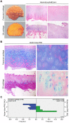
Histopathological features of OA articular cartilage. (A) Representative macroscopic images of fractured and OA femoral head samples (left panel) and Haematoxylin&Eosin-stained sections of articular cartilage at low and high magnification (center and right panel). (B) Representative images at different magnification of Alcian blue-PAS staining and (C) alcianophilia in the extracellular matrix of articular cartilage of fractured (n=30 donors; blue bars) and OA patients (n=30 donors; green bars). Abbreviations: A.U., arbitrary units. Mann-Whitney’s U-test: P < 0.0001.
Figure 2
Figure 2
IL-6 and AcH4 expression in articular cartilage of fractured and OA patients. (A) Representative immunohistochemical images of IL-6 and AcH4 expression in femoral cartilage of fractured (n=30) and OA patients (n=30). (B) Bar graphs show semi-quantitative evaluation of IL-6 and AcH4. Results are expressed as mean values ± SEM. Student’s t-test: *P < 0.001.
Figure 3
Figure 3
Morphology and proliferation rate of chondrocytes from OA and fractured patients in vitro. (A) Phase contrast micrographs of human chondrocytes in culture from articular cartilage of fractured and OA patients (F-HACs and OA-HACs). (B) Growth curve of F-HACs and OA-HACs in culture. (C) Representative images of Ki67 and p21 immunocytochemistry on F-HACs and OA-HACs. Bar graphs show the percentage of Ki67+ and p21+ cells. Data are expressed as mean values ± SEM of three independent experiments. Abbreviation: F-HACs, fractured patient human articular chondrocytes; OA-HACs, OA-Human articular chondrocytes. Student’s t-test: *P < 0.05, **P < 0.001, ***P < 0.0001.
Figure 4
Figure 4
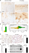
CLU expression in cartilage and chondrocyte cultures of fractured and OA patients. (A) Representative images of CLU immunohistochemistry on femoral cartilage of fractured and OA patients. (B) Percentage of CLU+ cells and (C) extracellular CLU secretion in femoral cartilage of fractured (n=30 donors; yellow bars) and OA patients (n=30 donors; green bars). Student’s t-test: **P < 0.001 and Mann-Whitney’s U-test: P < 0.0001, respectively. (D) Representative images and semi-quantitative evaluation of CLU immunocytochemistry on cytospinned HACs from fractured and OA patients. Data are expressed as mean values ± SEM of three independent experiments (each using pooled samples from n=3 fractured and n=3 OA donors) performed in triplicate. (E) CLU mRNA levels by Real-Time PCR and (F) representative blot and densitometric analysis of CLU protein in cultured HACs from fractured and OA cartilage of individual donors (n=10 fractured and n=10 OA patients). Experiments were performed in triplicate. Data are expressed as mean values ± SEM. Abbreviations: A.U, arbitrary units; F-HACs, fractured patient human articular chondrocytes; OA-HACs, OA-Human articular chondrocytes; psCLU, CLU precursor protein. Student’s t-test: *P < 0.01, ***P < 0.0001.
Figure 5
Figure 5
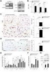
Effects of CLU silencing on viability, proliferation/death and differentiation of cultured OA chondrocytes. (A) CLU mRNA and protein expression after 48 and 72 hours, respectively, in scramble (control) and siCLU transfected OA-HACs. (B) Cell viability by MTT assay after 24, 48 and 72 hours of scramble and siCLU transfected OA-HACs. (C) Representative immunocytochemical images and bar graphs showing Ki67+ and p21+ nuclei in cultured OA-HACs and (D) cleaved caspase-3+ cytospinned OA-HACs after 72 hours of silencing. (E) Gene array shows the modulation of NFkB and Stat-3-related genes in siCLU transfected OA-HACs compared with scramble after 48 hours of silencing. Transcript levels were normalized on GAPDH (internal reference gene). (F) mRNA levels by Real-Time PCR of COL2A1, ACAN, MMP13, COL10A1, and (G) representative blot and densitometric analysis of MMP13 and COL10A1 in scramble and CLU-silenced OA-HACs. Data are expressed as mean values ± SEM of three independent experiments (each using pooled samples from n=3 OA donors) performed in triplicate. Abbreviations: OA-HACs, OA-Human articular chondrocytes; OD, optical density unit; RLU, relative light unit. Student’s t-test: *P < 0.05, **P < 0.01, ***P < 0.001, ****P < 0.0001.
Figure 6
Figure 6
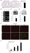
Effects of CLU silencing on TNFα and ROS levels in cultured OA chondrocytes. (A) Representative immunocytochemical images and evaluation of TNFα+ cytospinned OA-HACs after 72 hours of CLU silencing. (B) Basal mRNA expression of NOX isoforms by RT-PCR in cultured chondrocytes. (C, D) RT-PCR and blot analysis of NOX4 expression in scramble and CLU-silenced OA-HACs. (E, F) Representative images and quantitative measurement of ROS levels (green fluorescence) after 12, 24 and 48 hours of CLU silencing compared with control (scramble) and HACs from fractured patients; original magnification, 100X. Data are expressed as mean values ± SEM of three independent experiments (each using pooled samples from n=3 donors) performed in triplicate. Abbreviations: F-HACs, fractured patient human articular chondrocytes; OA-HACs, OA-Human articular chondrocytes; ADU, arbitrary densitometric unit; F.I., fluorescence intensity. Student’s t-test: *P < 0.05, **P < 0.01 and ***P < 0.001.

References

    1. Zhang Y, Jordan JM. Epidemiology of osteoarthritis. Clin Geriatr Med. 2010; 26:355–69. 10.1016/j.cger.2010.03.001 - DOI - PMC - PubMed
    1. Dieppe PA, Lohmander LS. Pathogenesis and management of pain in osteoarthritis. Lancet. 2005; 365:965–73. 10.1016/S0140-6736(05)71086-2 - DOI - PubMed
    1. Johnson VL, Hunter DJ. The epidemiology of osteoarthritis. Best Pract Res Clin Rheumatol. 2014; 28:5–15. 10.1016/j.berh.2014.01.004 - DOI - PubMed
    1. Goldring MB. The role of the chondrocyte in osteoarthritis. Arthritis Rheum. 2000; 43:1916–26. 10.1002/1529-0131(200009)43:9<1916::AID-ANR2>3.0.CO;2-I - DOI - PubMed
    1. Hinton R, Moody RL, Davis AW, Thomas SF. Osteoarthritis: diagnosis and therapeutic considerations. Am Fam Physician. 2002; 65:841–48. - PubMed

Publication types

MeSH terms