. 2020 Sep 1;130(9):4935-4946.
doi: 10.1172/JCI134965.

PRICKLE3 linked to ATPase biogenesis manifested Leber's hereditary optic neuropathy

Affiliations

PRICKLE3 linked to ATPase biogenesis manifested Leber's hereditary optic neuropathy

Jialing Yu et al. J Clin Invest. .

Abstract

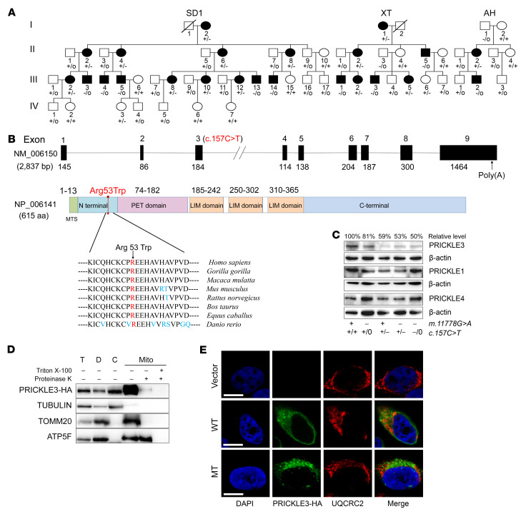
Leber's hereditary optic neuropathy (LHON) is a maternally inherited eye disease. X-linked nuclear modifiers were proposed to modify the phenotypic manifestation of LHON-associated mitochondrial DNA (mtDNA) mutations. By whole-exome sequencing, we identified the X-linked LHON modifier (c.157C>T, p.Arg53Trp) in PRICKLE3 encoding a mitochondrial protein linked to biogenesis of ATPase in 3 Chinese families. All affected individuals carried both ND4 11778G>A and p.Arg53Trp mutations, while subjects bearing only a single mutation exhibited normal vision. The cells carrying the p.Arg53Trp mutation exhibited defective assembly, stability, and function of ATP synthase, verified by PRICKLE3-knockdown cells. Coimmunoprecipitation indicated the direct interaction of PRICKLE3 with ATP synthase via ATP8. Strikingly, cells bearing both p.Arg53Trp and m.11778G>A mutations displayed greater mitochondrial dysfunction than those carrying only a single mutation. This finding indicated that the p.Arg53Trp mutation acted in synergy with the m.11778G>A mutation and deteriorated mitochondrial dysfunctions necessary for the expression of LHON. Furthermore, we demonstrated that Prickle3-deficient mice exhibited pronounced ATPase deficiencies. Prickle3-knockout mice recapitulated LHON phenotypes with retinal deficiencies, including degeneration of retinal ganglion cells and abnormal vasculature. Our findings provided new insights into the pathophysiology of LHON that were manifested by interaction between mtDNA mutations and X-linked nuclear modifiers.

Keywords: Genetic diseases; Genetics; Mitochondria; Molecular pathology; Ophthalmology.


Conflict of interest statement

Conflict of interest: The authors have declared that no conflict of interest exists.

Figures

Figure 1
Figure 1. Identification of PRICKLE3 p.Arg53Trp mutation.
(A) Three Chinese pedigrees with LHON. Vision-impaired individuals are indicated by blackened symbols. Individuals harboring hemizygous (–/0) or heterozygous (+/–) PRICKLE3 (c.157C>T, p.Arg53Trp) mutation and WT (+/+ or +/0) are indicated. (B) Scheme for the structure of PRICKLE3 and its product, multiple sequence alignment of homologs. (C) Western blot analysis of PRICKLE3, PRICKLE1, and PRICKLE4 in various lymphoblastoid cell lines. The levels of these proteins were quantified in the linear range of the sample loading and expressed as percentages of the average values for the control cell lines. (D) Subcellular localization of PRICKLE3 by Western blotting with anti-HA, TOMM20 (outer mitochondrial membrane), ATP5F (inner mitochondrial membrane), and tubulin (cytosol). T, total cell lysate; D, debris; C, cytosol; Mito, mitochondria. Isolated mitochondria were treated with (+) or without (–) 1% Triton X-100 followed by proteinase K digestion, respectively. (E) Subcellular localization of PRICKLE3 by immunofluorescence in HeLa cells. HA-PRICKLE3 WT or mutant (MT) (shown in green), UQCRC2 (shown in red), and DAPI (shown in blue). Scale bar: 10 μm. See complete unedited blots in the supplemental material.
Figure 2
Figure 2. The p.Arg53Trp mutation caused the defective activity of mitochondrial ATPase.
(A) Enzymatic activities of respiratory chain complexes in mitochondria isolated from various cell lines. (B) In-gel activity of ATPase. (C) The rates of OCR (O2 consumption) in the various cell lines using different inhibitors. Basal OCR was determined as OCR before oligomycin minus OCR after rotenone/antimycin A. ATP-lined OCR was determined as OCR before oligomycin minus OCR after oligomycin. (D) Measurement of mitochondrial ATP levels using a bioluminescence assay. Cells were incubated with 5 mM 2-deoxy-d-glucose plus 5 mM pyruvate to determine ATP generation under mitochondrial ATP synthesis. Average rates of ATP level per cell line and are shown. Dashed lines indicate the mean values of enzymatic activities (A), OCRs (C), or ATP production (D) in PRICKLE3+/– and PRICKLE3–/0 cell lines. Data are shown as mean ± SEM. *P < 0.05; **P < 0.01; ***P < 0.001 by 2-way ANOVA (A and C) and 1-way ANOVA (D) followed by Bonferroni’s post hoc test; NS, not significant. Representative of 3 to 4 independent experiments.
Figure 3
Figure 3. The p.Arg53Trp mutation altered the assembly and stability of ATP synthase.
(A and B) The stability of fully assembled ATP synthase monomer. Mitochondria isolated from various lymphoblastoid cell lines (A) and HeLa cells, which were transfected by shRNA (PRICKLE3 and scramble) and rescued by WT-PRICKLE3 (B), subjected to blue native PAGE, and hybridized with anti-ATP5A antibody and VDAC as an internal control. Data are shown as mean ± SEM of triplicates. (C) Immunoblot analysis of subunits of ATP synthase. Total cellular proteins in various lymphoblastoid cell lines were electrophoresed with PAGE and hybridized with ATP6, ATP8, ATPAF1, ATP5B, ATP5A, ATP5C, and ATP5F antibodies and VDAC as a loading control, respectively. (D) Physical interaction of PRICKLE3 with ATP8 (A6L). Isolated mitochondria from HEK293T cells, transiently expressing WT, MT PRICKLE3-HA, and empty vector, were solubilized with 0.5% DDM. Lysate proteins were immunoprecipitated with ATP synthase immunocapture kit (left), HA antibody (right), respectively. Antibodies: anti-HA and anti-ATP8, -ATP5A, -ATP5B, -ATP5F, and -ATPAF1 for complex V and anti-UQCRC2 for complex III were used, respectively. (E) Proposed model for the direct interaction between PRICKLE3 and ATPase (FO: a, b, 8/A6L, d, e, f, g, diabetes-associated protein in insulin-sensitive tissue; F1: α, β, γ, δ, ε). Matrix, mitochondrial matrix; IMM, inner mitochondrial membranes. FOF1 ATP structural model was modified from He et al. (37).
Figure 4
Figure 4. The altered activity and assembly of complex V in retinas from Prickle3-KO mice.
(A) Schema of generation of Prickle3-KO mice (C57BL/6J) using the CRISPR/Cas9 system. A 13-bp deletion was produced in exon 3 (c.144_158del) of Prickle3, resulting in a truncated protein with 60 amino acids. (B) Levels of Prickle3 in retinas from different genotypes of mice at 8 weeks of age. (C) Prickle3 expression in retinas of WT and Prickle3−/– mice by immunolabeling analysis. ONL, outer nuclear layer; OPL, outer plexiform layer; INL, inner nuclear layer; IPL, inner plexiform layer; RGL, ganglion cell layer. Scale bar: 10 μm. (D) Enzymatic activities of complexes I, II, III, IV, and V in mitochondria isolated from the livers of Prickle3-KO and WT mice. Dashed lines indicate the mean values of enzymatic activities in samples of Prickle3–/0 and Prickle3–/– mice. Data are shown as mean ± SEM of triplicates. ***P < 0.001 by 2-way ANOVA followed by Bonferroni’s post hoc test. (E) In-gel activity of ATPase in the livers of Prickle3-KO and WT mice. VM, complex V monomer. (F) Assembly of ATP synthase monomer using blue native PAGE analysis in the liver mitochondria of Prickle3-KO and WT mice. (G) Levels of ATP8, ATP6, ATP5A, and ATP5F in the retinas of Prickle3-KO and WT mice. (H) Mitochondrial morphology in RGCs by transmission electron microscopy. Arrows indicate mitochondria. Scale bars: 1 μm (upper panel), 0.2 μm (lower panel).
Figure 5
Figure 5. Retinal deficiencies in Prickle3-KO mice at postnatal 8 weeks.
(A) Retinal section staining with H&E in WT, Prickle3+/–, and Prickle3–/– mice. Arrows indicate RGC layer. Scale bar: 100 μm. (B) RGC staining with Brn3a (green) with β-III-tubulin (red) and DAPI (blue). The ratios of Brn3a+/DAPI+ cells in the RGC layer of WT (n = 5), Prickle3+/– (n = 5), and Prickle3–/– (n = 5) were 57% ± 8%, 46% ± 5%, and 36% ± 6% from 5 crossretinal sections for each genotypic mouse sample (lower panel), respectively. Scale bar: 20 μm. (C) Neurofilament staining of the whole-mount retina with anti–NF-H antibody and the neurons (NF positive) with their dendrites at higher magnification. Scale bars: 200 μm (upper panel), 50 μm (lower panel). (D) The mean dendritic areas measured from NF-H–positive cells in WT (n = 24), Prickle3+/– (n = 24), and Prickle3–/– (n = 24) genotypic mouse samples. Data are shown as mean ± SEM. ***P < 0.001 by 1-way ANOVA followed by Bonferroni’s post hoc test. (E) Expression of postsynaptic density-95 (Psd95) in WT, Prickle+/–, and Prickle3–/– retinas. (F) Retinal vasculature labeling with isolectin B4. Yellow arrow indicate the vessels in WT mice, and white arrows point to tortuous vessels in Prickle3+/– and Prickle3–/– mice. Scale bar: 200 μm. (G) Fluorescence angiography of eyes in WT, Prickle3+/–, and Prickle3–/– mice. (H) Analysis of ffERG for WT (n = 8), Prickle–/– (n = 4), or Prickle3–/0 (n = 4) mice. By dark adaptation for a night, mice were analyzed for scotopic response and then photopic response. Data are shown as mean ± SEM of triplicates. **P < 0.01; ***P < 0.001 by Student’s t test.

References

    1. Wallace DC, Lott MT. Leber hereditary optic neuropathy: exemplar of an mtDNA disease. Handb Exp Pharmacol. 2017;240:339–376. doi: 10.1007/164_2017_2. - DOI - PubMed
    1. Howell N. Leber hereditary optic neuropathy: respiratory chain dysfunction and degeneration of the optic nerve. Vision Res. 1998;38(10):1495–1504. doi: 10.1016/S0042-6989(97)00444-6. - DOI - PubMed
    1. Sadun AA, La Morgia C, Carelli V. Leber’s hereditary optic neuropathy. Curr Treat Options Neurol. 2011;13(1):109–117. doi: 10.1007/s11940-010-0100-y. - DOI - PubMed
    1. Newman NJ. Hereditary optic neuropathies: from the mitochondria to the optic nerve. Am J Ophthalmol. 2005;140(3):517–523. - PubMed
    1. Carelli V, La Morgia C, Valentino ML, Barboni P, Ross-Cisneros FN, Sadun AA. Retinal ganglion cell neurodegeneration in mitochondrial inherited disorders. Biochim Biophys Acta. 2009;1787(5):518–528. - PubMed

Publication types

MeSH terms