. 2020 Jun 3;61(6):20.
doi: 10.1167/iovs.61.6.20.

Role of TH17 Responses in Increasing Herpetic Keratitis in the Eyes of Mice Infected with HSV-1

Affiliations

Role of TH17 Responses in Increasing Herpetic Keratitis in the Eyes of Mice Infected with HSV-1

Satoshi Hirose et al. Invest Ophthalmol Vis Sci. .

Abstract

Purpose: TH17 cells play an important role in host defense and autoimmunity yet very little is known about the role of IL17 in herpes simplex virus (HSV)-1 infectivity. To better understand the relationship between IL17 and HSV-1 infection, we assessed the relative impact of IL17A-deficiency and deficiency of its receptors on HSV-1 responses in vivo.

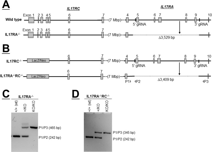
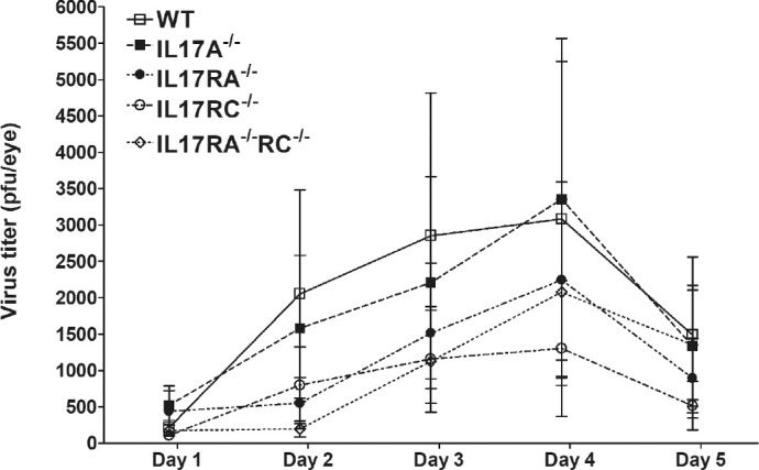
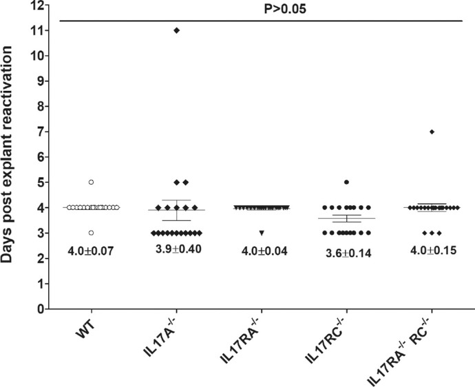
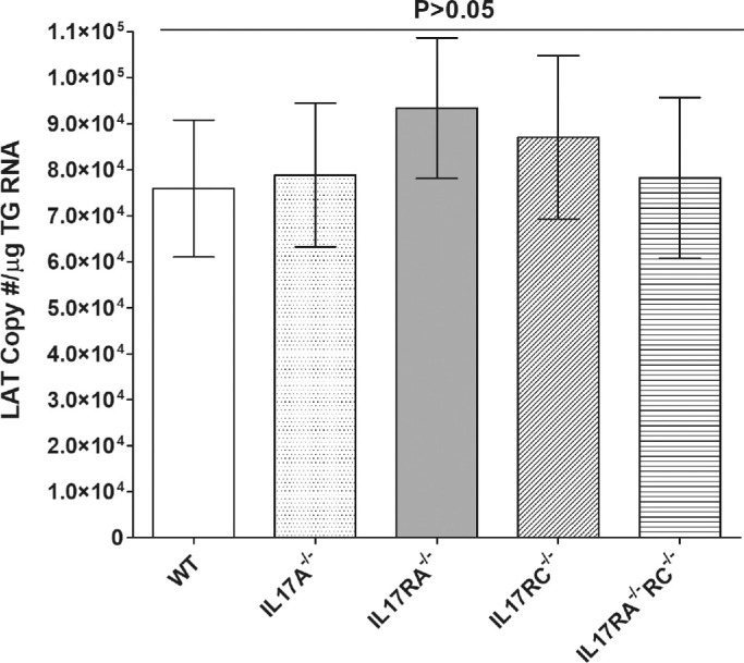
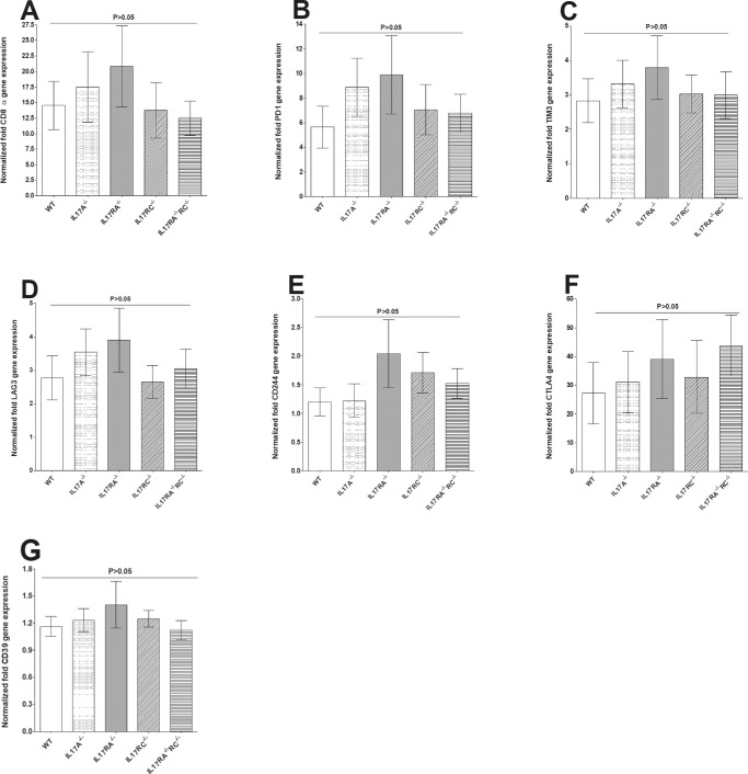
Methods: We generated IL17RA-/- and IL17RA-/-RC-/- mice in-house and infected them along with IL17A-/- and IL17RC-/- mice in the eyes with 2 × 10⁵ PFU/eye of wild type (WT) HSV-1 strain McKrae. WT C57BL/6 mice were used as control. Virus replication in the eye, survival, corneal scarring (CS), angiogenesis, levels of latency-reactivation, and levels of CD8 and exhaustion markers (PD1, TIM3, LAG3, CTLA4, CD244, and CD39) in the trigeminal ganglia (TG) of infected mice were determined on day 28 postinfection.

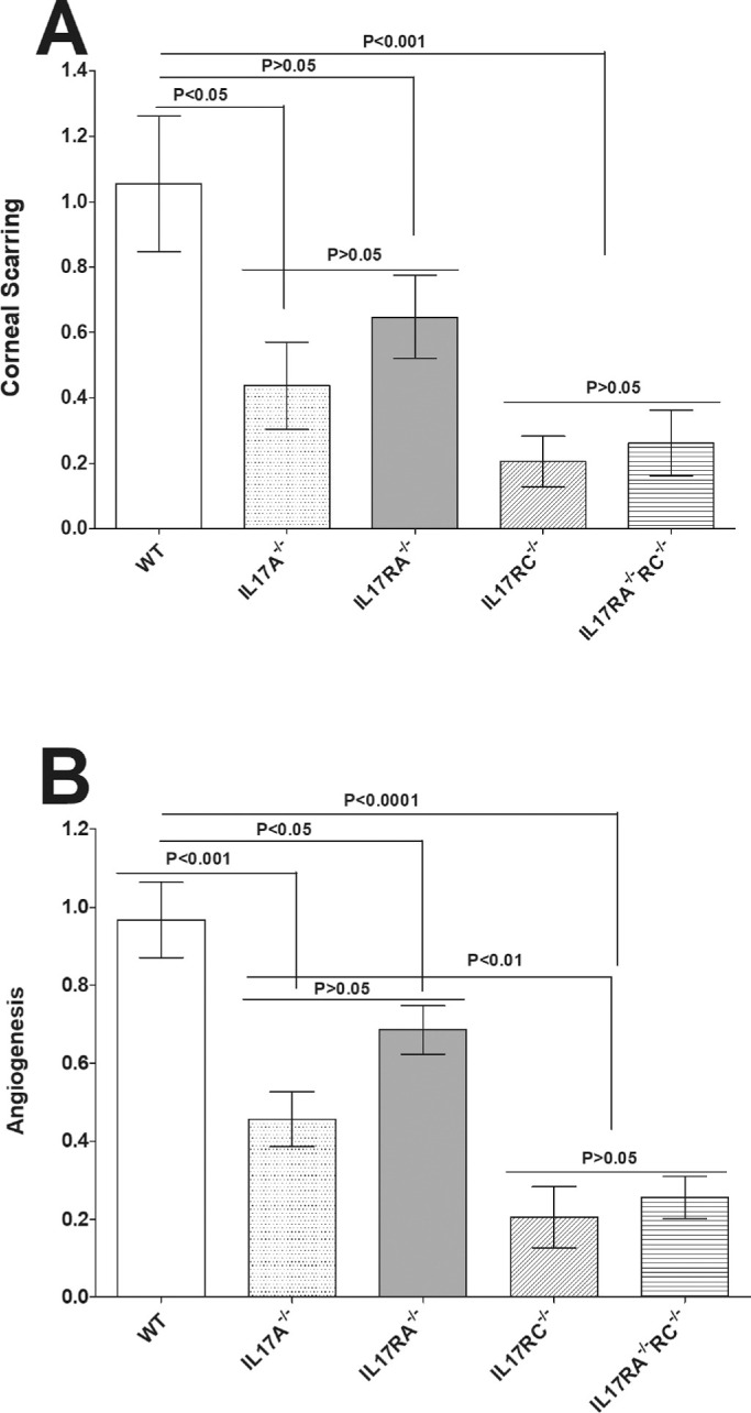
Results: No significant differences in virus replication in the eye, survival, latency, reactivation, and exhaustion markers were detected among IL17A-/-, IL17RA-/-, IL17RC-/-, IL17RA-/-RC-/-, and WT mice. However, mice lacking IL17 had significantly less CS and angiogenesis than WT mice. In addition, angiogenesis levels in the absence of IL17RC and irrespective of the absence of IL17RA were significantly less than in IL17A- or IL17RA-deficient mice.

Conclusions: Our results suggest that the absence of IL17 protects against HSV-1-induced eye disease, but has no role in protecting against virus replication, latency, or reactivation. In addition, our data provide rationale for blocking IL17RC function rather than IL17A or IL17RA function as a key driver of HSV-1-induced eye disease.


Conflict of interest statement

Disclosure: S. Hirose, None; U. Jaggi, None; S. Wang, None; K. Tormanen, None; Y. Nagaoka, None; M. Katsumata, None; H. Ghiasi, None

Figures

Figure 1.
Figure 1.
Generation of IL17RA−/− and IL17RA−/−RC−/− mice. (A) Schematic diagram of IL17RA−/− mice. Top diagram depicts genomic loci of IL17RC and IL17RA in wt mice. Vertical bars show binding sites of guide RNAs (gRNAs) used to target Cas 9 to exons 5 and 9 of IL17RA using wild type mice (exons are shown as gray boxes). Deletions generated by CRISPR method are shown as dotted lines in the bottom diagram; (B) Schematic diagram of IL17RA−/−RC−/− mice. Top diagram shows genomic loci of IL17RC and IL17RA in IL17RC−/− mice. Similar to A above, gRNAs were used to delete exons 5 to 9 of IL17RA in IL17RC−/− mice. Deletions generated by CRISPR method are shown as dotted lines in the bottom diagram, and P1, P2, and P3 indicate binding sites of primers used to detect deletions by PCR; (C) PCR to detect deletion of IL17RA in wt mice. DNA from mice generated in A was used to confirm deletion of IL17RA. PCR primers P1 and P2 yield the PCR product from wild type allele, while PCR primers P1 and P3 yield PCR products from the knockout alleles; and (D) PCR to detect deletion of IL17RA in IL17RC−/− mice. DNA from mice generated in B was used to confirm deletion of IL17RA in IL17RC−/− mice. PCR primers P1 and P2 yield the PCR product from wild type allele, while PCR primers P1 and P3 yield PCR products from the knockout alleles.
Figure 2.
Figure 2.
Virus titers in the eyes of infected mice. WT and IL17A−/−, IL17RA−/−, IL17RC−/−, and IL17RA−/−RC−/− mice were infected with 2 × 10⁵ PFU/eye of HSV-1 strain McKrae as described in the Materials and Methods. Tear films were collected on days 1-5 PI and virus titers were determined using standard plaque assays. Each point represents the mean ± SEM titers of 28 eyes for WT, 20 eyes for IL17A−/−; 26 eyes for IL17RA−/−; 26 eyes for IL17RC−/−; and 22 eyes for IL17RA−/−RC−/− mice from 2-4 separate experiments.
Figure 3.
Figure 3.
Loss of IL17 contributes to reduced eye disease. Corneal scarring (CS) and angiogenesis in surviving mice were assessed on day 28 PI as described in Materials and Methods. CS and angiogenesis were assessed in 46 eyes from WT, 48 eyes from IL17A−/− mice, 58 eyes from IL17RA−/− mice, 44 eyes from IL17RC−/− mice, and 44 eyes from IL17RA−/−RC−/− mice. Experiments were repeated three times and CS and angiogenesis scores are presented as mean ± SEM. p-value was determined using a one-way ANOVA test. Panels: A) CS in surviving mice; and B) Angiogenesis in surviving mice.
Figure 4.
Figure 4.
Duration of reactivation is not affected in IL17-deficient mice. To analyze explant reactivation in infected mice, WT and IL17A−/−, IL17RA−/−, IL17RC−/− and IL17RA−/−RC−/− mice were infected in the eye as described in Figure 2. On day 28 PI, TG from infected mice were harvested for explant reactivation. Each individual TG was incubated in 1.5 ml of tissue culture media at 37°C and the presence of infectious virus was monitored for 12 d. Reactivation is based on 21of 22, 20 of 26, 24 of 28,19 of 20, and 23 of 24 TG of WT and IL17A−/−, IL17RA−/−, IL17RC−/− and IL17RA−/−RC−/− mice, respectively. The average time that the TG from each group first showed CPE ± SEM is shown. p-value was determined using a one-way ANOVA test.
Figure 5.
Figure 5.
Latency levels in infected mice. To analyze levels of latency in the TG of latently-infected mice, WT and IL17A−/−, IL17RA−/−, IL17RC−/−, and IL17RA−/−RC−/− mice were infected in the eye as described in Figure 2 above. On day 28 PI, TG were harvested from the latently-infected mice. Quantitative RT-PCR was performed on individual TG from each mouse. GAPDH expression was used to normalize relative expression of LAT RNA in the TG. LAT copy number per TG were measured using pGEM5317, a LAT containing plasmid as we described previously. Latency is based on 18 TG per group of mice. p-value was determined using a one-way ANOVA test.
Figure 6.
Figure 6.
Roles of IL17 on CD8 and exhaustion markers in the TG of latently-infected mice. RNA isolated from WT and IL17A−/−, IL17RA−/−, IL17RC−/−, and IL17RA−/−RC−/− mice as described in Figure 5 were used to measure the expression of CD8α, PD1, TIM3, LAG3, CD244, CTLA4, and CD39 in latently-infected TG. qRT-PCR was performed using total RNA as described in the Materials and Methods. CD8α, PD1, TIM3, LAG3, CD244, CTLA4, and CD39 expression in naive WT mice was used as a baseline control to estimate relative expression of each transcript in TG of latently-infected mice. GAPDH expression was used to normalize the relative expression of each transcript. Each point represents the mean ± SEM from 10 to 12 TG for each strain of mice. p-value was determined using a one-way ANOVA test. Panels: A) CD8α transcript; B) PD1 transcript; C) TIM3 transcript; D) LAG3 transcript; E) CD244 transcript; F) CTLA4 transcript; and G) CD39 transcript.

References

    1. Sad S, Li L, Mosmann TR. Cytokine-deficient CD8+ Tc1 cells induced by IL-4: retained inflammation and perforin and Fas cytotoxicity but compromised long term killing of tumor cells. J Immunol. 1997; 159: 606–613. - PubMed
    1. Mosmann TR, Yokota T, Kastelein R, Zurawski SM, Arai N, Takebe Y. Species-specificity of T cell stimulating activities of IL 2 and BSF-1 (IL 4): comparison of normal and recombinant, mouse and human IL 2 and BSF-1 (IL 4). J Immunol. 1987; 138: 1813–1816. - PubMed
    1. Street NE, Schumacher JH, Fong TA, et al. .. Heterogeneity of mouse helper T cells. Evidence from bulk cultures and limiting dilution cloning for precursors of Th1 and Th2 cells. J Immunol. 1990; 144: 1629–1639. - PubMed
    1. Breitfeld D, Ohl L, Kremmer E, et al. .. Follicular B helper T cells express CXC chemokine receptor 5, localize to B cell follicles, and support immunoglobulin production. J Exp Med. 2000; 192: 1545–1552. - PMC - PubMed
    1. Schmitt E, Klein M, Bopp T. Th9 cells, new players in adaptive immunity. Trends Immunol. 2014; 35: 61–68. - PubMed

Publication types

MeSH terms