Review
. 2020 Jun;10(6):200051.
doi: 10.1098/rsob.200051. Epub 2020 Jun 10.

The centromere comes into focus: from CENP-A nucleosomes to kinetochore connections with the spindle

Affiliations
Review

The centromere comes into focus: from CENP-A nucleosomes to kinetochore connections with the spindle

Kathryn Kixmoeller et al. Open Biol. 2020 Jun.

Abstract

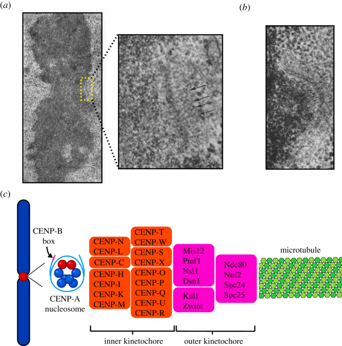
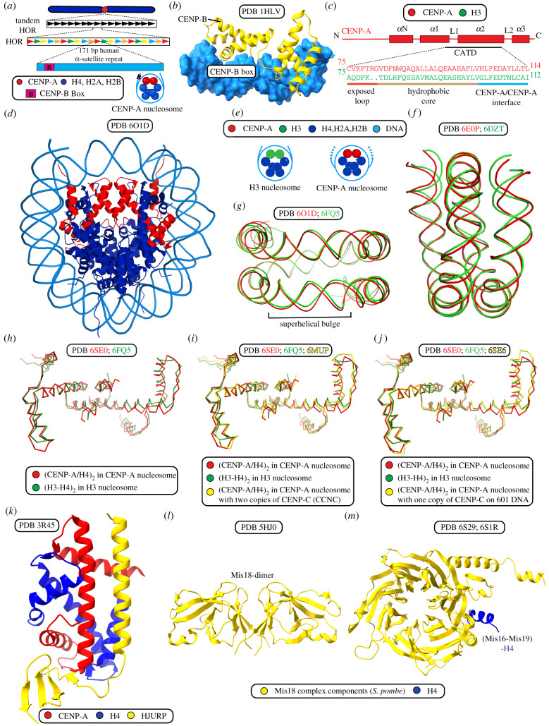
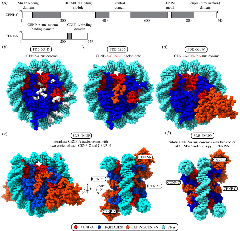
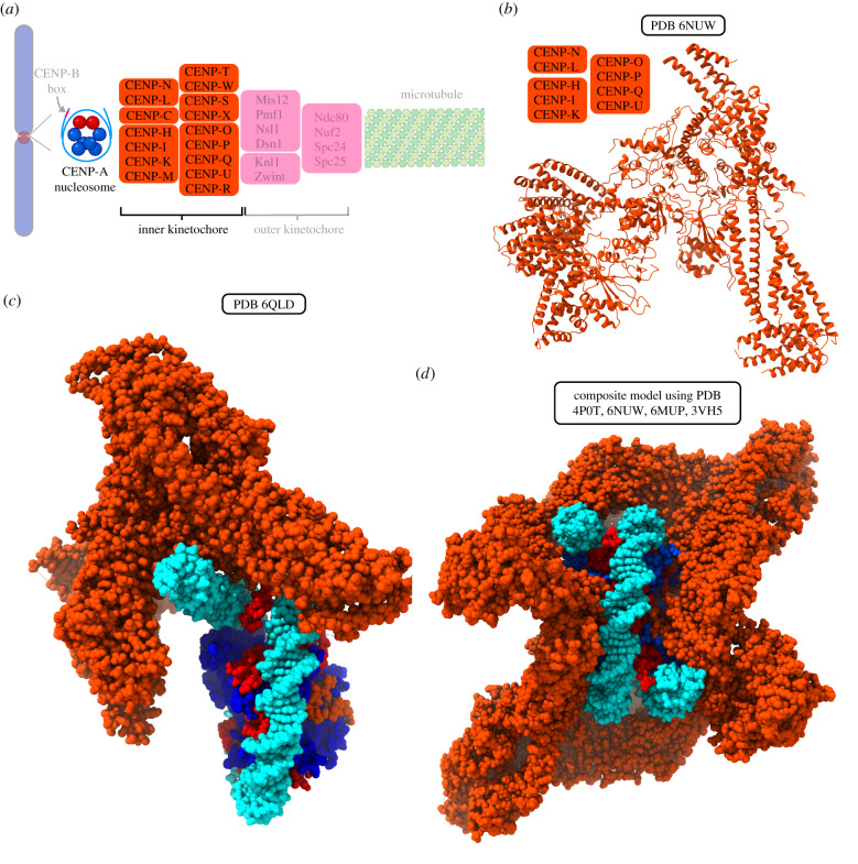
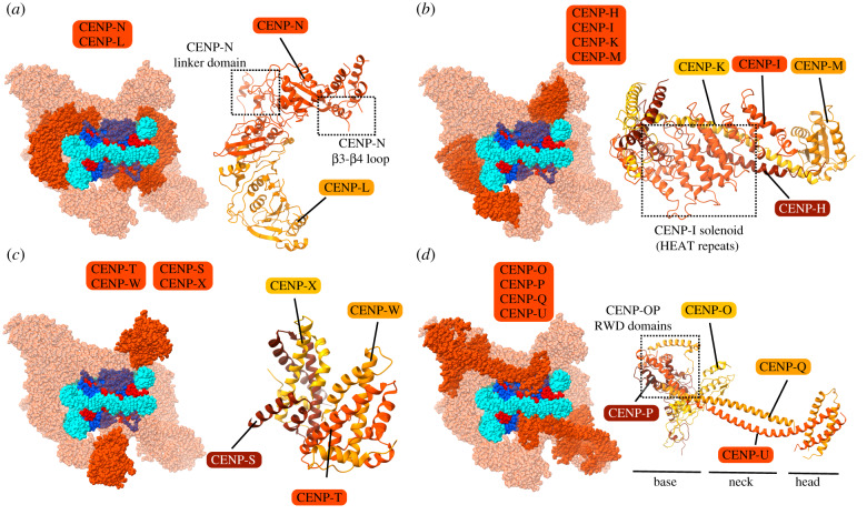
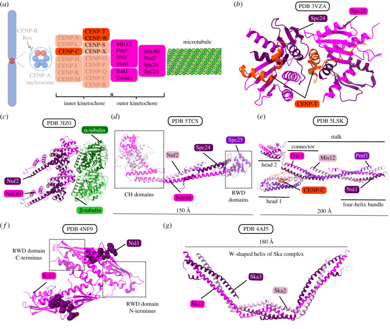
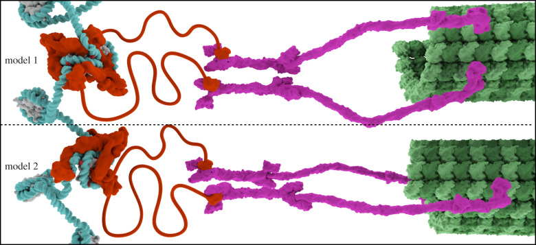
Eukaryotic chromosome segregation relies upon specific connections from DNA to the microtubule-based spindle that forms at cell division. The chromosomal locus that directs this process is the centromere, where a structure called the kinetochore forms upon entry into mitosis. Recent crystallography and single-particle electron microscopy have provided unprecedented high-resolution views of the molecular complexes involved in this process. The centromere is epigenetically specified by nucleosomes harbouring a histone H3 variant, CENP-A, and we review recent progress on how it differentiates centromeric chromatin from the rest of the chromosome, the biochemical pathway that mediates its assembly and how two non-histone components of the centromere specifically recognize CENP-A nucleosomes. The core centromeric nucleosome complex (CCNC) is required to recruit a 16-subunit complex termed the constitutive centromere associated network (CCAN), and we highlight recent structures reported of the budding yeast CCAN. Finally, the structures of multiple modular sub-complexes of the kinetochore have been solved at near-atomic resolution, providing insight into how connections are made to the CCAN on one end and to the spindle microtubules on the other. One can now build molecular models from the DNA through to the physical connections to microtubules.

Keywords: centromere; chromatin; epigenetics; kinetochore; mitosis; nucleosome.


Conflict of interest statement

We declare we have no competing interests.

Figures

Figure 1.
Figure 1.
Structural organization of the centromere and kinetochore. (a) Electron micrograph of a mitotic chromosome with paired kinetochores on either side of the primary constriction (centromere), prior to microtubule attachments [3]. The inset shows a higher-magnification electron micrograph, which further reveals the trilaminar structure of the kinetochore, showing fibrous elements on either side of the dense central kinetochore plate. Arrows indicate kinetochore fibrils extending out from centromere. Permission to reproduce was sought from the author, Dr William Brinkley, but no response was received. (b) This micrograph of the centromere–kinetochore region shows a crescent-shaped kinetochore and the fibrous corona extending outwards from it. (c) A schematic of the DNA-microtubule interface, from the CENP-A nucleosomes found in centromeric DNA, through the many subunits of the kinetochore complex, and finally to the microtubule.
Figure 2.
Figure 2.
Structure and assembly of CENP-A chromatin. (a) Human centromeres typically are located within 0.5–5 Mb of α-satellite DNA arranged in large higher-order repeats (HOR) where the smallest repeating unit is 171 bp. The CENP-B box is located within the human 171 bp α-satellite centromeric repeat monomer, mostly outside of the CENP-A nucleosome DNA entry/exit site. (b) The structure of the human CENP-B N-terminal domain as bound to CENP-B box DNA. (c) Alignment of the CENP-A targeting domain (CATD) with the corresponding region of canonical histone H3. The CENP-A targeting domain (CATD) provides a distinct surface and features to centromeric CENP-A nucleosomes when compared with canonical H3 nucleosomes. The CATD provides three crucial characteristics: (1) CENP-A/CENP-A interface rotation; (2) strong interactions at the CENP-A/H4 interface due to hydrophobic stitch residues; (3) a protruding loop L1, which gives CCAN specificity) that combine to make the centromeric CENP-A nucleosome distinct. (d) Structure of a CENP-A nucleosome assembled on 145 bp human α-satellite DNA. (e) Schematic of DNA entry/exit dynamics. CENP-A nucleosomes show precise positioning on 171 bp α-satellite repeat and show flexibility in the terminal DNA predicted by access to MNase digestion. H3 nucleosomes lack these features when assembled on the same repeats. These features are important for the assembly of kinetochore proteins and are specific for CENP-A nucleosomes due to shortened α-1 helix in CENP-A. (f) DNA flexibility within the CENP-A nucleosome impacts the path of terminal DNA in all available cryo-EM and X-ray crystal CENP-A nucleosome structures relative to H3 nucleosome structures. The differences in the paths of DNA bound to CENP-A (red) and H3 (green) nucleosomes can be observed by alignment of the nucleosome cryo-EM structures using the (CENP-A/H4)2 and (H3/H4)2 dimers, as shown here. (g) A distinct feature of CENP-A nucleosome structures assembled on α-satellite DNA is the presence of a superhelical bulge, which is absent from H3 nucleosome structures and from CENP-A nucleosome structures assembled on 601 DNA or palindromic α-satellite DNA sequences. The superhelical bulge in the path of the nucleosomal DNA can be observed here by the alignment of CENP-A nucleosomes assembled on α-satellite DNA (red) with H3 nucleosomes assembled on 601 DNA (green) using the CENP-A/H4 dimer and H3/H4 dimer. The superhelical bulge of CENP-A nucleosomes provides a surface for accurate assembly of CCAN proteins. (h–j) Illustrations of the overall impact of intrinsic features of CENP-A and DNA sequence on nucleosome structure by alignment of the CENP-A/H4 dimer to the H3/H4 dimer or the CENP-A/H4 dimer, in the presence or absence of CENP-C from available structures. (h) CENP-A (red) and H3 (green) nucleosomes on 601 DNA. (i) CENP-A (red) and H3 (green) nucleosome on 601 DNA and CENP-A (yellow) nucleosome with CENP-C on α-satellite DNA. (j) CENP-A (red), H3 (green) nucleosome and CENP-A with CENP-C nucleosome (yellow) on 601 DNA. (k) The structure of the human CENP-A/H4 dimer in complex with its chaperone, HJURP. (l) Crystal structure of the CENP-A nucleosome assembly regulator, S. pombe Mis18. The structure includes its N-terminal Yippee-like domain, which is known to act as centromere targeting domain and contains a cradle-shaped pocket which binds DNA and is required for Mis18 functions. (m) Structure of the CENP-A nucleosome assembly regulatory complex member, S. pombe Mis16 with histone H4.
Figure 3.
Figure 3.
The CCNC forms the foundation of the kinetochore. (a) Domain coordinates for human CENP-C and CENP-N. (b) The CENP-A nucleosome structure is shown facing the surface of the histone octamer, with the dyad at top. White positions highlight the points on the protein and DNA surface where CCNC components have direct contacts. (c) The CENP-A nucleosome assembled with Widom 601 DNA and bound with the central domain of CENP-C. (d) The CENP-A nucleosomes assembled with Widom 601 DNA and bound with CENP-N. (e) The CENP-A nucleosome assembled with human α-satellite DNA and bound with two copies of CENP-C and CENP-N, proposed to be the interphase form of the CCNC. (f) The CENP-A nucleosome assembled with human α-satellite DNA and bound with two copies of CENP-C and one copy of CENP-N, proposed to be the mitotic form of the CCNC.
Figure 4.
Figure 4.
Structure of the yeast CCAN and models of the human CCAN complex bound to CENP-A nucleosome. (a) A schematic of the DNA–microtubule interface, with the CENP-A nucleosome and inner kinetochore highlighted. (b) Structure of the S. cerevisiae Ctf19/CCAN complex containing homologues to human CENP-LN, CENP-HIK and CENP-OPQUR. (c) Cryo-EM structure of one copy of S. cerevisiae Ctf19 (lacking homologues of CENP-M and CENP-TWSX) interacting with CENP-A nucleosome on Widom 601 DNA. In this structure, CENP-NChl14 does not contact CENP-A or proximal DNA at the sites described in structures of mammalian CENP-N. (d) Composite model of CCAN (Ctf19 complex/CENP-M/CENP-TWSX) with the CCNC. Structural alignment was performed by aligning the N-terminal domain of the common subunit, CENP-N.
Figure 5.
Figure 5.
Organization of CCAN sub-complexes on CENP-A nucleosomes. (ad) Location of indicated complex within the composite model of CCAN-CCNC complex (left) and ribbon diagram in isolation (right).
Figure 6.
Figure 6.
The KMN complex and other microtubule couplers. (a) A schematic of the DNA-microtubule interface, highlighting the microtubule, outer kinetochore and components of the inner kinetochore that couple the KMN to CENP-A nucleosomes. (b) Crystal structure of chicken Spc24/Spc25 globular domain (part of the Ndc80 complex) bound with CENP-T. This interaction allows up to two Ndc80 complexes to be recruited for each copy of CENP-T. (c) Structure of the S. cerevisiae ‘bonsai’ chimeric Ndc80 complex bound to tubulin. The ‘bonsai’ structure contains minimal coiled-coil domains, and Ndc80 is fused to Spc25 and Nuf2 to Spc24. The Ndc80 complex binds microtubules via N-terminal calponin homology domains in Ndc80 and Nuf2. (d) Structure of the dwarf Ndc80 tetramer, which is shortened with respect to the full complex but maintains the full tetramer junction. (e) Crystal structure of the human Mis12 complex bound with a fragment of the CENP-C N-terminal region. The Mis12 complex also interacts with CENP-T, Knl1 and Ndc80, serving as an important interaction hub between the KMN assembly and the inner kinetochore. (f) Crystal structure of the C-terminal RWD domains of human Knl1 interacting with Nsl1 residues. Knl1 is the largest structural subunit in the outer kinetochore and provides binding sites for Nsl1 as well as several proteins that interact with the outer kinetochore. (g) Crystal structure of the human Ska complex. The Ska complex functions to enhance the microtubule binding ability of the Ndc80 complex.
Figure 7.
Figure 7.
Models of the kinetochore organization on centromeric chromatin. Model 1 has a symmetric arrangement of two copies of the CCAN bound per CENP-A nucleosome, whereas model 2 has an asymmetric arrangement with a single copy of the CCAN per CENP-A nucleosome. Both models contain two copies of CENP-C per CENP-A nucleosome. The proposed connection through CENP-C to neighbouring nucleosomes has been omitted from both models for visual clarity. See text for details about the two models, the data supporting each one and implications for centromeric chromatin structure during mitotic chromosome segregation.

References

    1. Rieder CL, Salmon ED. 1998. The vertebrate cell kinetochore and its roles during mitosis. Trends Cell Biol. 8, 310–318. (10.1016/S0962-8924(98)01299-9) - DOI - PMC - PubMed
    1. Lampson MA, Grishchuk EL. 2017. Mechanisms to avoid and correct erroneous kinetochore-microtubule attachments. Biology 6, 1 (10.3390/biology6010001) - DOI - PMC - PubMed
    1. Brinkley BR, Stubblefield E. 1966. The fine structure of the kinetochore of a mammalian cell in vitro. Chromosoma 19, 28–43. (10.1007/BF00332792) - DOI - PubMed
    1. Cooke CA, Bazett-Jones DP, Earnshaw WC, Rattner JB. 1993. Mapping DNA within the mammalian kinetochore. J. Cell Biol. 120, 1083–1091. (10.1083/jcb.120.5.1083) - DOI - PMC - PubMed
    1. Musacchio A, Desai A. 2017. A molecular view of kinetochore assembly and function. Biology 6, 5 (10.3390/biology6010005) - DOI - PMC - PubMed

Publication types

LinkOut - more resources