. 2020 Jul 2;107(1):158-163.
doi: 10.1016/j.ajhg.2020.05.013. Epub 2020 Jun 8.

Mutations in ASPRV1 Cause Dominantly Inherited Ichthyosis

Affiliations

Mutations in ASPRV1 Cause Dominantly Inherited Ichthyosis

Lynn M Boyden et al. Am J Hum Genet. .

Abstract

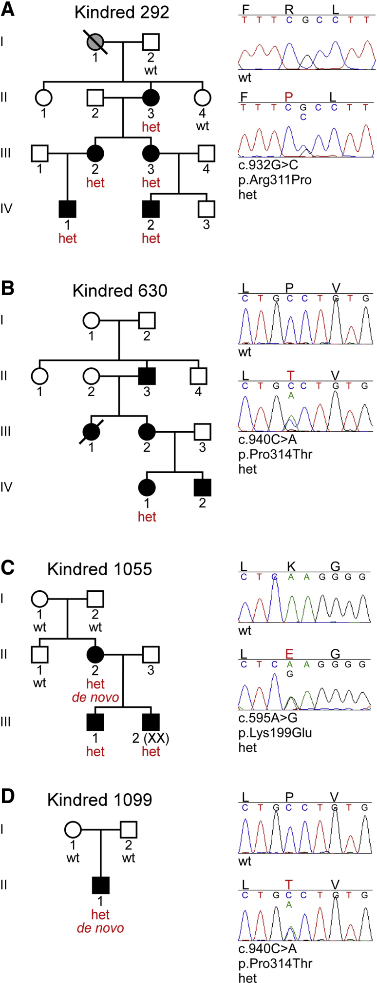
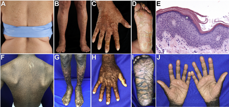
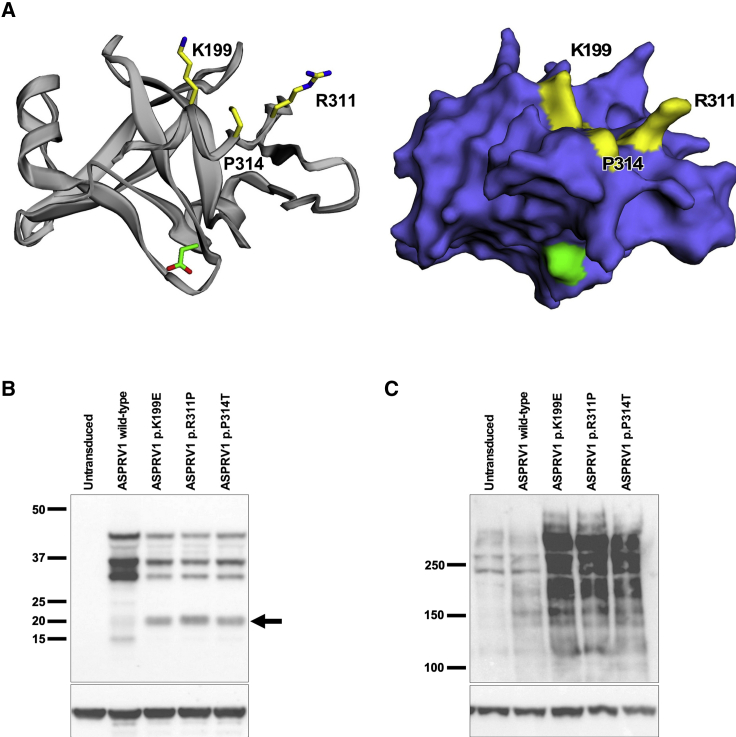
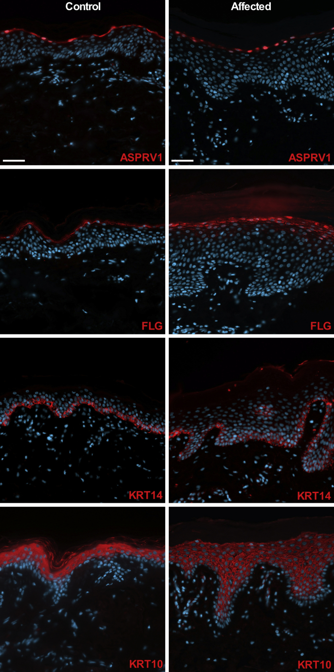
The discovery of genetic causes of inherited skin disorders has been pivotal to the understanding of epidermal differentiation, function, and renewal. Here we show via exome sequencing that mutations in ASPRV1 (aspartic peptidase retroviral-like 1) cause a dominant Mendelian disorder featuring palmoplantar keratoderma and lamellar ichthyosis, a phenotype that has otherwise been exclusively recessive. ASPRV1 encodes a mammalian-specific and stratified epithelia-specific protease important in processing of filaggrin, a critical component of the uppermost epidermal layer. Three different heterozygous ASPRV1 missense mutations in four unrelated ichthyosis kindreds segregate with disease and disrupt protein residues within close proximity to each other and autocatalytic cleavage sites. Expression of mutant ASPRV1 proteins demonstrates that all three mutations alter ASPRV1 auto-cleavage and filaggrin processing, a function vital to epidermal barrier integrity.

Keywords: ASPRV1; Mendelian; SASPase; de novo; dominant; epidermis; exome; ichthyosis; keratoderma; skin.


Figures

Figure 1
Figure 1
Ichthyosis Subjects Heterozygous for ASPRV1 Mutations Sanger sequencing was performed subsequent to exome sequencing results (Table S1). (A) In kindred 292, five subjects (II-3, III-2, III-3, IV-1, IV-2) are heterozygous (het) for c.932G>C, encoding p.Arg311Pro; two unaffected first-degree relatives (I-2, II-4) are wild-type (WT) at this site. (B) In kindred 630, the proband (IV-1) is heterozygous for c.940C>A, encoding p.Pro314Thr. DNA from other members of the kindred was not available. (C) In kindred 1055, the proband (III-1) and his affected mother (II-2) and brother (III-2) are heterozygous for c.595A>G, encoding p.Lys199Glu. This mutation is absent from the unaffected maternal uncle (II-1) and grandparents (I-1, I-2), and thus arose de novo in the proband’s mother. (D) In kindred 1099, the proband (II-1) is heterozygous for c.940C>A, encoding p.Pro314Thr, which is absent from his unaffected parents (I-1, I-2) and thus arose de novo.
Figure 2
Figure 2
Subjects Heterozygous for ASPRV1 Mutations Display a Consistent Skin Phenotype (A–E) Subject 292-2. There are large, plate-like, geometric scales over the back and posterior arms (A) and shins (B), corrugated hyperkeratosis over the extensor wrist and dorsal hand (C), and hyperlinearity of the foot, with large, plate-like, adherent scale and a few fissures of the heel (D). Histology (E) shows acanthosis, compact orthohyperkeratosis, and a prominent stratum granulosum with coarse keratohyalin granules containing filaggrin (asterisk). (F–J) Subject 1099. There is similar scale on the back (F), with larger plate-like scale on the lower extremities and feet (G), corrugated hyperkeratosis and smoothened scale over the hand (H), marked hyperlinearity of the feet with thick hyperkeratosis and many deep fissures (I), and hyperlinearity and hyperkeratosis of the palms with evidence of constricting bands of hyperkeratosis (pseudoainhum) on multiple digits (J).
Figure 3
Figure 3
Clustered ASPRV1 Mutations Generate a Novel ASPRV1 Auto-cleavage Product and Result in Accumulation of High-Molecular-Weight Forms of Filaggrin (A) A model of the ASPRV1 active enzyme crystal structure (ribbon and stick rendering on left, surface rendering on right) shows that all three pathogenic mutation sites (yellow) are tightly clustered within the three-dimensional protein. The catalytic aspartate is shown in green. (B and C) Human keratinocytes (HK), either nontransduced or transduced with wild-type or mutant ASPRV1, were switched to high calcium (1.2 mM) for 8 days to promote differentiation. Immunoblotting of protein extracts was performed with anti-actin (Sigma SAB4200248, 1:2,000) as a loading control on replicate blots to show equivalent protein loading across all samples. (B) Immunoblotting with an anti-HA antibody (Cell Signaling 3724, 1:500) shows bands at ~37 kDa and ~28 kDa in cells expressing either wild-type or mutant forms of HA-tagged ASPRV1. In addition, cells expressing each of the ASPRV1 mutant forms show a unique ~20 kDa band not seen in nontransduced cells or cells expressing wild-type ASPRV1 (arrow). (C) Immunoblotting with an anti-filaggrin antibody directed against the N-terminal domain (residues 1–261, Thermo Fisher Scientific PA5-79267, 1:500) shows profilaggrin and higher molecular weight filaggrin cleavage products within differentiated keratinocytes. In nontransduced keratinocytes and keratinocytes transduced with wild-type ASPRV1, there is a low quantity of high-molecular-weight filaggrin products, consistent with normal processing of filaggrin to filaggrin repeat monomers. Cells expressing mutant ASPRV1 (encoding p.Lys199Glu, p.Arg311Pro, or p.Pro314Thr) demonstrate an accumulation of high-molecular-weight filaggrin products, suggesting that ASPRV1 mutations impair filaggrin processing.
Figure 4
Figure 4
ASPRV1 Mutant Skin Shows Expansion of Filaggrin Immunolocalization ASPRV1 is localized to the cytoplasm and nuclei of cells within the stratum granulosum in age-matched control skin and affected tissue from subject 292-2. In contrast to the tightly focused filaggrin (FLG) immunolocalization noted in control tissue, affected tissue shows strong signal within a thickened granular layer of the epidermis and weak expression in the upper layers of stratum spinosum. Immunolocalization of keratin 14 (KRT14, a basal keratinocyte marker) is restricted to the basal layer and keratin 10 (KRT10, a suprabasal keratinocyte marker) is found in suprabasal layers in control and affected tissue. Scale bars are 50 μm. Primary antibodies (red staining) are rabbit anti-ASPRV1 (Sigma HPA034809, 1:200), rabbit anti-FLG (BioLegend 905801, 1:200), mouse anti-KRT14 (Santa Cruz sc-53252, 1:200), and guinea pig anti-KRT10 (Origene AP09544SU-N, 1:300). Nuclei are stained with DAPI (blue staining).

References

    1. Adzhubei I.A., Schmidt S., Peshkin L., Ramensky V.E., Gerasimova A., Bork P., Kondrashov A.S., Sunyaev S.R. A method and server for predicting damaging missense mutations. Nat. Methods. 2010;7:248–249. - PMC - PubMed
    1. Traupe H., Kolde G., Happle R. Autosomal dominant lamellar ichthyosis: a new skin disorder. Clin. Genet. 1984;26:457–461. - PubMed
    1. Kolde G., Happle R., Traupe H. Autosomal-dominant lamellar ichthyosis: ultrastructural characteristics of a new type of congenital ichthyosis. Arch. Dermatol. Res. 1985;278:1–5. - PubMed
    1. Bernard D., Méhul B., Thomas-Collignon A., Delattre C., Donovan M., Schmidt R. Identification and characterization of a novel retroviral-like aspartic protease specifically expressed in human epidermis. J. Invest. Dermatol. 2005;125:278–287. - PubMed
    1. Rhiemeier V., Breitenbach U., Richter K.H., Gebhardt C., Vogt I., Hartenstein B., Fürstenberger G., Mauch C., Hess J., Angel P. A novel aspartic proteinase-like gene expressed in stratified epithelia and squamous cell carcinoma of the skin. Am. J. Pathol. 2006;168:1354–1364. - PMC - PubMed

Publication types

Substances