. 2020 Sep;585(7824):273-276.
doi: 10.1038/s41586-020-2423-5. Epub 2020 Jun 9.

Clinical benefit of remdesivir in rhesus macaques infected with SARS-CoV-2

Affiliations

Clinical benefit of remdesivir in rhesus macaques infected with SARS-CoV-2

Brandi N Williamson et al. Nature. 2020 Sep.

Abstract

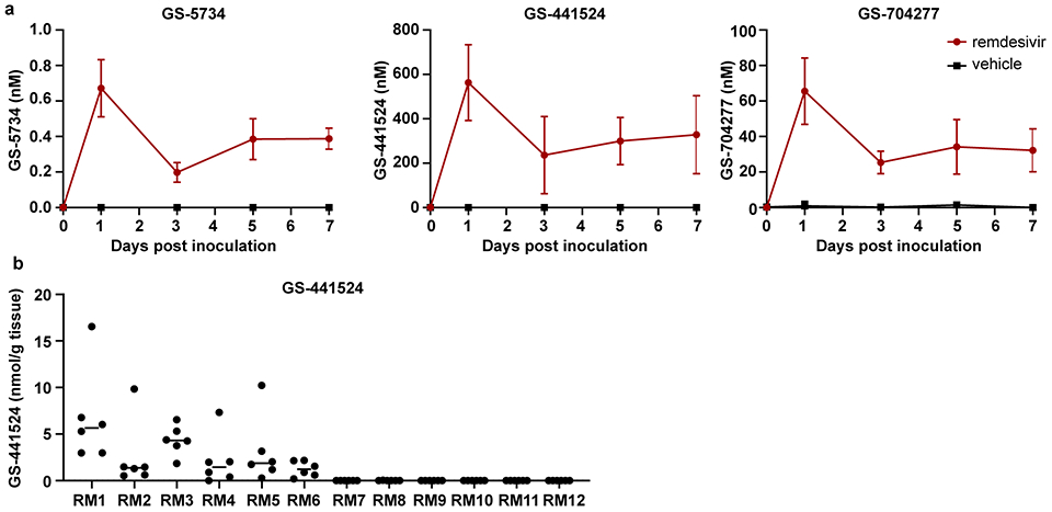
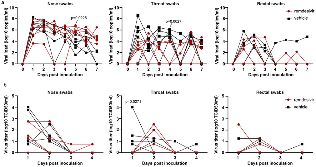
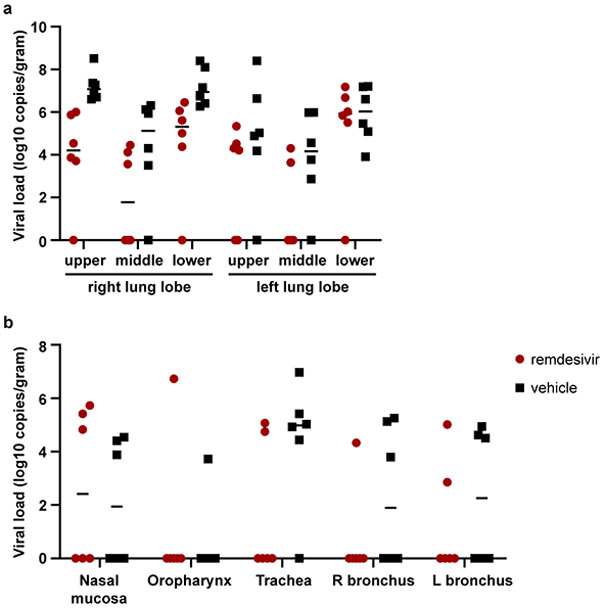
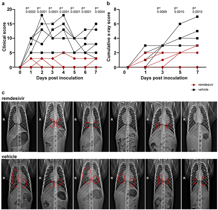
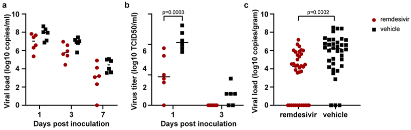
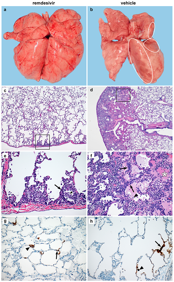
Effective therapies to treat coronavirus disease 2019 (COVID-19) are urgently needed. While many investigational, approved, and repurposed drugs have been suggested as potential treatments, preclinical data from animal models can guide the search for effective treatments by ruling out those that lack efficacy in vivo. Remdesivir (GS-5734) is a nucleotide analogue prodrug with broad antiviral activity1,2 that is currently being investigated in COVID-19 clinical trials and recently received Emergency Use Authorization from the US Food and Drug Administration3,4. In animal models, remdesivir was effective against infection with Middle East respiratory syndrome coronavirus (MERS-CoV) and severe acute respiratory syndrome coronavirus (SARS-CoV)2,5,6. In vitro, remdesivir inhibited replication of SARS-CoV-27,8. Here we investigate the efficacy of remdesivir in a rhesus macaque model of SARS-CoV-2 infection9. Unlike vehicle-treated animals, macaques treated with remdesivir did not show signs of respiratory disease; they also showed reduced pulmonary infiltrates on radiographs and reduced virus titres in bronchoalveolar lavages twelve hours after the first dose. Virus shedding from the upper respiratory tract was not reduced by remdesivir treatment. At necropsy, remdesivir-treated animals had lower lung viral loads and reduced lung damage. Thus, treatment with remdesivir initiated early during infection had a clinical benefit in rhesus macaques infected with SARS-CoV-2. Although the rhesus macaque model does not represent the severe disease observed in some patients with COVID-19, our data support the early initiation of remdesivir treatment in patients with COVID-19 to prevent progression to pneumonia.


Conflict of interest statement

Conflict of interest

The authors affiliated with Gilead Sciences are employees of the company and own company stock. The authors affiliated with NIH have no conflict of interest to report.

Figures

Extended Data Figure 1.
Extended Data Figure 1.. Concentration of remdesivir prodrug and metabolites measured in serum and lung homogenates of rhesus macaques infected with SARS-CoV-2.
Two groups of six rhesus macaques were inoculated with SARS-CoV-2 strain nCoV-WA1-2020. Twelve hours post inoculation, one group was administered 10mg/kg intravenous remdesivir and the other group was treated with an equal volume of vehicle solution (2ml/kg). Treatment was continued 12hrs after the first treatment, and every 24 hrs thereafter with a dose of 5 mg/kg remdesivir or equal volume of vehicle solution (1ml/kg). (a) Serum concentration of remdesivir prodrug GS-5734, the dephosphorylated nucleoside product GS-441524 and the intermediate alanine metabolite GS-704277 over time as measured in by LCMS for all animals (n=12) in the study. Mean and standard deviation are shown. (b) Concentration of GS-441524 homogenized lung tissue collected from all six lung lobes from each animal (n=12) on 7 dpi, 24 hrs after the last remdesivir treatment was administered. Each dot represents the concentration of GS-441524 in one lung lobe. The center bar represents the median.
Extended Data Figure 2.
Extended Data Figure 2.. Viral loads and virus titers in swabs collected from rhesus macaques infected with SARS-CoV-2 and treated with remdesivir.
Panel A shows viral loads and Panel B shows infectious virus titers in nose, throat and rectal swabs collected daily from animals treated with remdesivir (n=6) or vehicle solution (n=6). Statistical analysis was performed using a 2-way ANOVA with Sidak’s multiple comparisons test.
Extended Data Figure 3.
Extended Data Figure 3.. Viral loads in tissues collected from the respiratory tract on 7 dpi.
(a) Viral loads in all six lung lobes collected from rhesus macaques infected with SARS-CoV-2 and treated with remdesivir (n=6) or vehicle solution (n=6), stratified per lung lobe. (b) Viral loads in other tissues collected throughout the respiratory tract on 7 dpi. The center bar represents the median.
Extended Data Figure 4.
Extended Data Figure 4.. Pathological changes in lungs of rhesus macaques infected with SARS-CoV-2 and treated with remdesivir.
Rhesus macaques infected with SARS-CoV-2 and treated with remdesivir (n=6) or vehicle solution (n=6) were euthanized on 7 dpi. (a) The area of each individual lung lobe affected by gross lesions as scored by a veterinary pathologist blinded to group assignment of the animals. (b) All data from panel a combined. (c) Lung weight: bodyweight ratio as an indicator of pulmonary edema. (d) cumulative histology score. Each lung lobe was scored for the presence of histologic lung lesions on a predetermined scale (0-4); these values were combined per animal and graphed. Data in panel a were analyzed using a 2-way ANOVA with Sidak’s multiple comparisons test; data in panels b-d were analyzed using a two-tailed, unpaired t test. The center bar represents the median.
Figure 1.
Figure 1.. Reduced respiratory disease in rhesus macaques infected with SARS-CoV-2 and treated with remdesivir.
(a) Daily clinical scores in animals infected with SARS-CoV-2 and treated with remdesivir (red circles, n=6) or vehicle solution (black squares, n=6). (b) Cumulative radiograph scores. Ventro-dorsal and lateral radiographs were scored for the presence of pulmonary infiltrates by a clinical veterinarian according to a standard scoring system (0: normal; 1: mild interstitial pulmonary infiltrates; 2: moderate pulmonary infiltrates perhaps with partial cardiac border effacement and small areas of pulmonary consolidation; 3: severe interstitial infiltrates, large areas of pulmonary consolidation, alveolar patterns and air bronchograms). Individual lobes were scored and scores per animal per day were totaled and displayed. (c) Ventro-dorsal radiographs collected from each animal taken on 7 dpi. Areas of pulmonary infiltration are marked with a circle. Statistical analysis was performed using a 2-way ANOVA with Sidak’s multiple comparisons test.
Figure 2.
Figure 2.. Viral loads and virus titers in bronchoalveolar lavage fluid and lung lobes.
(a) Viral loads and (b) infectious virus titers in BAL collected from rhesus macaques infected with SARS-CoV-2 and treated with remdesivir (n=6) or vehicle solution (n=6). Statistical analysis was performed using a 2-way ANOVA with Sidak’s multiple comparisons test. (c) Viral loads in tissues collected from all six lung lobes at necropsy on 7 dpi from rhesus macaques infected with SARS-CoV-2 and treated with remdesivir (n=6) or vehicle solution (n=6). Statistical analyses was performed using an unpaired t test. The center bars in each panel indicate the median.
Figure 3.
Figure 3.. Changes to the lungs of rhesus macaques infected with SARS-CoV-2 and treated with remdesivir.
Rhesus macaques infected with SARS-CoV-2 and treated with remdesivir (n=6) or vehicle solution (n=6) were euthanized on 7 dpi. (a) Representative dorsal view of lungs of a remdesivir-treated animal. (b) Representative dorsal view of lungs of a vehicle-treated animal with focally extensive areas of consolidation (circles). Histological analysis was performed on 3 sections from 6 lung lobes from each of the 6 animals per treatment group and representative images were chosen for panels c-h. (c) Minimal subpleural interstitial pneumonia (box) observed in 3 of 6 remdesivir-treated animals. (d) Moderate subpleural interstitial pneumonia with edema (box) observed in 5 of 6 vehicle-treated animals. (e) Boxed area from panel c with alveoli lined by type II pneumocytes (arrow) and alveolar spaces containing foamy macrophages (arrowhead). (f) Boxed area from panel d with pulmonary interstitium expanded by edema and moderate numbers of macrophages and neutrophils. Alveoli are lined by type II pneumocytes (arrows). Alveolar spaces are filled with edema (asterisk) and small numbers of pulmonary macrophages (arrowhead). (g) Viral antigen in type I pneumocytes (arrow) and type II pneumocytes (arrowhead) of a remdesivir-treated animal. (h) Viral antigen in type I pneumocytes (arrow) and macrophage (arrowhead) of a vehicle-treated animal. Magnification c and d: 40x; panel e-h: 200x.

Update of

References

    1. Lo MK, Jordan R, Arvey A, et al. GS-5734 and its parent nucleoside analog inhibit Filo-, Pneumo-, and Paramyxoviruses. Sci Rep 2017;7:43395. - PMC - PubMed
    1. Sheahan TP, Sims AC, Graham RL, et al. Broad-spectrum antiviral GS-5734 inhibits both epidemic and zoonotic coronaviruses. Sci Transl Med 2017;9. - PMC - PubMed
    1. Remdesivir. 2020. (Accessed 05/15/2020, at https://clinicaltrials.gov/ct2/results?cond=&term=remdesivir&cntry=&stat....)
    1. Coronavirus (COVID-19) Update: FDA Issues Emergency Use Authorization for Potential COVID-19 Treatment. 2020. (Accessed 05/15/2020, at https://www.fda.gov/news-events/press-announcements/coronavirus-covid-19....)
    1. de Wit E, Feldmann F, Cronin J, et al. Prophylactic and therapeutic remdesivir (GS-5734) treatment in the rhesus macaque model of MERS-CoV infection. Proc Natl Acad Sci U S A 2020. - PMC - PubMed

Publication types

MeSH terms